Monoclonal Antibodies
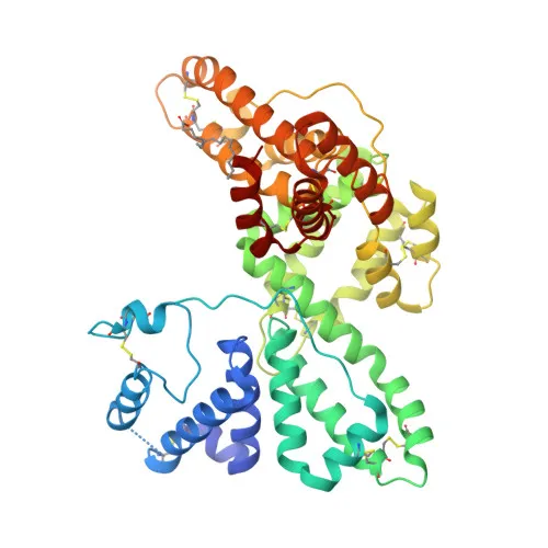
Anti-Vitamin D-binding protein Rabbit Monoclonal Antibody for WB, ELISA - P02774
Item Number : CM0003669
Price varies based on specs and customizations
- Application
- WB, ELISA
- Cross Reactivity
- Human, Mouse, Rat
- Protein Weight
- 53kDa

Shipping:
Contact us to get shipping details Enjoy On-time Dispatch Guarantee.
Why Choose Us
Easy ordering process, quality products, and dedicated support for your business success.
Core Product Specifications and Parameters
| Parameter | Value |
|---|---|
| Product Name | Vitamin D Binding protein Rabbit mAb |
| Remarks/Alias | DBP; VDB; GRD3; VDBG; VDBP; GcMAF; DBP/GC; Gc-MAF; DBP-maf; HEL-S-51; Vitamin D Binding protein |
| Species | Human |
| GeneID (Human) | 2638 |
| GeneID | 2638 |
| Immunogen | Recombinant fusion protein containing a sequence corresponding to amino acids 305-474 of human Vitamin D Binding protein (NP_000574.2) |
| Source | Rabbit |
| Category | Monoclonal Antibodies |
| Application | WB, ELISA |
| Cross Reactivity | Human, Mouse, Rat |
| SWISS | P02774 |
| Protein Weight | 53kDa |
| Shipping | Ice bag |
Biological Background: Vitamin D-binding protein Function and Localization
- Vitamin D-binding protein (DBP), also known as Gc-globulin or group-specific component (Gc), is a multifunctional plasma protein primarily synthesized in the liver and secreted into the bloodstream.
- It plays a central role in vitamin D transport and storage, binding vitamin D and its metabolites, thereby ensuring their solubility and systemic distribution.
- DBP acts as an extracellular actin scavenger, binding to globular G-actin released upon cell injury to prevent actin polymerization and potential damage to the microcirculation.
- The protein enhances the chemotactic activity of complement component C5a for neutrophils, contributing to inflammation and innate immune responses.
- Alternative splicing generates isoforms, and post-translational modifications include glycosylation and disulfide bond formation, which can influence its function and stability.
- DBP is not only found in plasma but also in other body fluids such as ascites, cerebrospinal fluid, and urine, reflecting its wide distribution and potential localized roles.
Experimental Guidance and Technical Tips
- The immunogen corresponds to the C-terminal region (amino acids 305-474) of human DBP, which encompasses parts of the molecule involved in vitamin D and actin binding; this design may allow recognition of both full-length and certain cleavage products.
- For Western blot, detect DBP at approximately 53 kDa, but be aware that glycosylation and other modifications may cause slight shifts in observed molecular weight.
- Cross-reactivity with mouse and rat DBP is predicted; however, sequence alignment should be checked to confirm epitope conservation, and pilot experiments are recommended when using these species.
- For ELISA, consider the potential presence of DBP variants or bound ligands in samples that could affect antibody binding; appropriate controls are advised.
CamelBio: Your One-Stop Sourcing Bridge
CamelBio streamlines IVD raw-material procurement by providing diagnostic manufacturers and research laboratories with validated antibody pairs, optimized monoclonal/polyclonal antibodies, and bulk ancillary reagents. For targets like Vitamin D-binding protein — critical in vitamin D metabolism, immune regulation, and inflammation — we offer tailored sourcing solutions, including rare-target raw materials, to support your assay development from concept to clinic. Partner with CamelBio to accelerate your diagnostic innovation with reliable, high-performance reagents.
Product Datasheet
Anti-Vitamin D-binding protein Rabbit Monoclonal Antibody for WB, ELISA - P02774
REQUEST A QUOTE
Our professional team will reply to you within one business day. Please feel free to contact us!