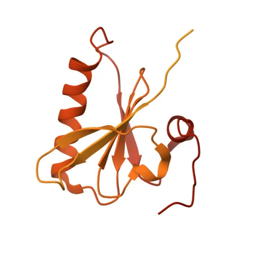
Monoclonal Antibodies
Anti-Villin Monoclonal Antibody for WB, IHC-P, ELISA - P09327
Item Number : CM0004774
Price varies based on specs and customizations
- Application
- WB, IHC-P, ELISA
- Cross Reactivity
- Human, Mouse, Rat
- Protein Weight
- 93kDa

Shipping:
Contact us to get shipping details Enjoy On-time Dispatch Guarantee.
Why Choose Us
Easy ordering process, quality products, and dedicated support for your business success.
Core Product Specifications and Parameters
| Parameter | Value |
|---|---|
| Product Name | Villin Rabbit mAb |
| Remarks/Alias | VIL; D2S1471; Villin |
| Species | Human |
| Gene ID | 7429 |
| Immunogen | Recombinant fusion protein containing a sequence corresponding to amino acids 1-827 of human Villin1 (NP_009058.2) |
| Source | Rabbit |
| Category | Monoclonal Antibodies |
| Application | WB, IHC-P, ELISA |
| Cross Reactivity | Human, Mouse, Rat |
| SWISS | P09327 |
| Protein Weight | 93kDa |
| Shipping | Ice bag |
Biological Background: Villin Function and Localization
- Villin-1 (VIL1) is an epithelial cell-specific actin-modifying protein that regulates microvillar actin filament reorganization; alternative splicing produces multiple isoforms.
- It bundles, caps, severs, and nucleates actin filaments, playing a central role in the dynamics of the actin cytoskeleton in brush border microvilli.
- Activity is tightly regulated by Ca²⁺ and phosphoinositides: PIP₂ inhibits capping and severing but enhances bundling, while LPA binding promotes SRC-mediated phosphorylation and inhibits all actin-modifying functions.
- Subcellularly, villin localizes to the cytoplasm, cytoskeleton, and actin-rich cell projections including microvilli, filopodia tips, lamellipodia, and ruffles.
- Tissue expression is highly restricted to epithelial cells; it is a major component of microvilli in intestinal epithelium, kidney proximal tubule cells, and canalicular microvilli of hepatocytes.
- Beyond cytoskeletal remodeling, villin regulates cell morphology, invasion, migration, and apoptosis; it protects against DSS-induced epithelial apoptosis by maintaining mitochondrial integrity.
- Upon Shigella flexneri infection, villin's actin-severing activity facilitates bacterial dissemination by enhancing actin-based motility.
- Additional keywords include actin capping, actin-binding, apoptosis, calcium, cell projection, phosphoprotein, and repeat domains, reflecting its multifunctional nature.
Experimental Guidance and Technical Tips
- For Western blot, a single band at approximately 93 kDa is expected in epithelial lysates; consider using intestinal or kidney tissue extracts as positive controls.
- In immunohistochemistry (paraffin), verify staining specificity in human, mouse, or rat intestinal sections due to high villin expression in brush borders.
- The immunogen spans nearly full-length villin (aa 1–827), suggesting recognition of multiple epitopes; validate detection in your specific sample system if working with splice variants or fragments.
- For ELISA, recombinant villin protein can serve as the coating antigen; titration of the antibody is recommended to optimize the signal-to-noise ratio.
- Cross-reactivity with mouse and rat makes this antibody suitable for studies in rodent models; however, confirm performance under your experimental conditions.
CamelBio: Your One-Stop Sourcing Bridge
CamelBio is your integrated partner for IVD raw materials, streamlining procurement of validated monoclonal and polyclonal antibodies like this anti-Villin clone for epithelial and cytoskeletal research. We supply optimized antibody pairs, bulk ancillary reagents, and offer customized sourcing for rare targets, supporting diagnostic manufacturers and labs from assay development to clinical application. Let CamelBio bridge your material needs with reliable, one-stop access to quality reagents.
REQUEST A QUOTE
Our professional team will reply to you within one business day. Please feel free to contact us!