Polyclonal Antibodies
Anti-VDR Rabbit Polyclonal Antibody for WB, IF/ICC, ELISA - P11473
Item Number : CM0021551
Price varies based on specs and customizations
- Application
- WB, IF/ICC, ELISA
- Cross Reactivity
- Human, Mouse
- Protein Weight
- 48kDa

Shipping:
Contact us to get shipping details Enjoy On-time Dispatch Guarantee.
Why Choose Us
Easy ordering process, quality products, and dedicated support for your business success.
Core Product Specifications and Parameters
| Parameter | Value |
|---|---|
| Product Name | VDR Rabbit pAb |
| Remarks/Alias | NR1I1; PPP1R163; VDR |
| Species | Human |
| GeneID | 7421 |
| Immunogen | Synthetic peptide|A synthetic peptide corresponding to a sequence within amino acids 1-100 of human VDR (NP_000367.1). |
| Source | Rabbit |
| Category | Polyclonal Antibodies |
| Application | WB, IF/ICC, ELISA |
| Cross Reactivity | Human, Mouse |
| SWISS | P11473 |
| Protein Weight | 48kDa |
| Shipping | Ice bag |
Biological Background: VDR Function and Localization
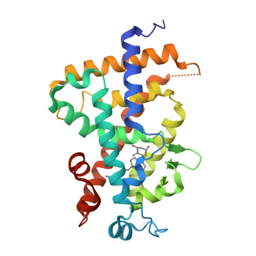
- VDR (Vitamin D3 receptor), also known as NR1I1 (nuclear receptor subfamily 1 group I member 1) and PPP1R163, is a member of the nuclear receptor family of transcription factors.
- It functions as the receptor for calcitriol (1,25-dihydroxyvitamin D3), the active form of vitamin D, mediating genomic actions upon ligand binding. Related references: PMID:10678179, PMID:15728261, PMID:16913708
- Upon vitamin D3 binding, VDR translocates to the nucleus and forms heterodimers with the retinoid X receptor (RXR) to regulate transcription of target genes. Related references: PMID:28698609
- VDR plays a central role in calcium homeostasis by controlling the expression of genes involved in calcium absorption and bone metabolism.
- Beyond its classical endocrine role, VDR also functions as a receptor for secondary bile acid lithocholic acid (LCA) and its metabolites, linking vitamin D signaling to bile acid detoxification. Related references: PMID:12016314, PMID:32354638
- The protein localizes to both the nucleus and cytoplasm, with nuclear translocation enhanced upon ligand activation.
- Key structural features include a DNA-binding domain with zinc-finger motifs and a ligand-binding domain, as indicated by keywords: zinc, zinc-finger, DNA-binding, metal-binding.
- Alternative splicing generates multiple isoforms, and the protein is subject to post-translational modifications including ubiquitin conjugation (Ubl conjugation).
Experimental Guidance and Technical Tips
- The immunogen corresponds to a synthetic peptide from the N-terminal region (amino acids 1–100) of human VDR, potentially recognizing both full-length and certain variant isoforms.
- For Western blot (WB), consider using whole-cell lysates from VDR-expressing tissues or cell lines; note the predicted molecular weight of 48 kDa.
- In immunofluorescence/immunocytochemistry (IF/ICC), optimize fixation and permeabilization steps to detect both cytoplasmic and nuclear VDR signals.
- For ELISA applications, validate the antibody as a capture or detection reagent with recombinant VDR protein or peptide standards.
- Always include appropriate controls (e.g., VDR-overexpressing lysates, blocking peptide) to confirm specificity in your experimental system.
CamelBio: Your One-Stop Sourcing Bridge
CamelBio provides diagnostic manufacturers, laboratories, and research institutes with one-stop access to IVD raw materials, technical services, and consulting—supporting projects from concept to clinic. For VDR-related research and assay development, we offer a broad portfolio including validated antibody pairs, bulk ancillary reagents, and custom sourcing for rare targets. Contact us to learn how our integrated supply solutions can streamline your vitamin D receptor studies and diagnostic kit production.
REQUEST A QUOTE
Our professional team will reply to you within one business day. Please feel free to contact us!