Polyclonal Antibodies
Anti-VAV1 Rabbit Polyclonal Antibody for WB, ELISA - P15498
Item Number : CM0013715
Price varies based on specs and customizations
- Application
- WB, ELISA
- Cross Reactivity
- Human, Mouse, Rat
- Protein Weight
- 98kDa

Shipping:
Contact us to get shipping details Enjoy On-time Dispatch Guarantee.
Why Choose Us
Easy ordering process, quality products, and dedicated support for your business success.
Core Product Specifications and Parameters
| Parameter | Value |
|---|---|
| Product Name | VAV1 Rabbit pAb |
| Remarks/Alias | VAV; VAV1 |
| Species | Human |
| GeneID | 7409 |
| Immunogen | Recombinant fusion protein containing a sequence corresponding to amino acids 374-590 of human VAV1 (NP_005419.2) |
| Source | Rabbit |
| Category | Polyclonal Antibodies |
| Application | WB, ELISA |
| Cross Reactivity | Human, Mouse, Rat |
| SWISS | P15498 |
| Protein Weight | 98kDa |
| Shipping | Ice bag |
Biological Background: VAV1 Function and Localization
- VAV1 encodes the proto-oncogene vav, a member of the VAV family of guanine nucleotide exchange factors (GEFs) that activate Rho/Rac GTPases.
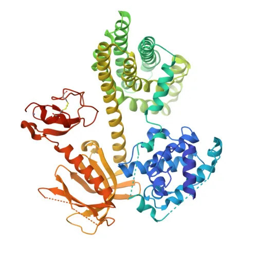
- The protein contains multiple conserved domains, including SH2, SH3, a zinc-finger domain, and a Dbl homology domain essential for its GEF activity.
- VAV1 serves as a critical signal transducer that links tyrosine kinase signaling to the activation of Rho/Rac GTPases, thereby promoting cell differentiation, proliferation, and cytoskeletal reorganization.
- The protein is selectively expressed in hematopoietic cells, including lymphocytes and myeloid lineages, whereas it is generally absent in non‑hematopoietic tissues.
- VAV1 undergoes alternative splicing and is regulated by phosphorylation at multiple tyrosine residues, modulating its GEF activity and interaction with downstream partners.
- Zinc‑ and metal‑binding motifs within VAV1 are integral to its structural stability and catalytic function as a guanine‑nucleotide releasing factor.
Experimental Guidance and Technical Tips
- The immunogen, spanning amino acids 374–590 of human VAV1, encompasses the SH3 domain and adjacent regions that are conserved in mouse and rat; this design likely underpins the antibody’s multi‑species reactivity.
- For Western blotting, expect a band near 98 kDa. To validate specificity, use lysates from hematopoietic cell lines (e.g., Jurkat, THP‑1) and verify absence in non‑hematopoietic cells, given the tissue‑restricted expression of VAV1.
- In ELISA applications, optimize coating antigen concentration and antibody dilution; the polyclonal nature may provide robust signal but requires careful titration to minimize background.
- Owing to VAV1’s hematopoietic specificity, experimental designs should include appropriate positive controls (e.g., activated T‑cells) and negative controls (e.g., fibroblast lines) to confirm on‑target detection.
CamelBio: Your One-Stop Sourcing Bridge
CamelBio empowers diagnostic manufacturers, laboratories, and research institutes with comprehensive, one‑stop access to IVD raw materials, including validated antibody pairs, optimized monoclonal and polyclonal antibodies, bulk ancillary reagents, and rare‑target sourcing. Our VAV1 rabbit polyclonal antibody exemplifies our commitment to supporting hematopoietic signaling and oncology research with high‑quality reagents. As your sourcing bridge, we streamline procurement from early‑stage development to clinical application, ensuring you have the critical components needed to advance your IVD projects.
REQUEST A QUOTE
Our professional team will reply to you within one business day. Please feel free to contact us!