Polyclonal Antibodies
Anti-VASH1 Polyclonal Antibody for WB, ELISA - Q7L8A9
Item Number : CM0024932
Price varies based on specs and customizations
- Application
- WB, ELISA
- Cross Reactivity
- Human, Mouse
- Protein Weight
- 41kDa

Shipping:
Contact us to get shipping details Enjoy On-time Dispatch Guarantee.
Why Choose Us
Easy ordering process, quality products, and dedicated support for your business success.
Core Product Specifications and Parameters
| Parameter | Value |
|---|---|
| Product Name | VASH1 Rabbit pAb |
| Remarks/Alias | TTCP 1; KIAA1036; VASH1 |
| Species | Human |
| Gene ID (Human) | 22846 |
| Gene ID | 22846 |
| Immunogen | Recombinant fusion protein containing a sequence corresponding to amino acids 1-365 of human VASH1 (NP_055724.1) |
| Source | Rabbit |
| Category | Polyclonal Antibodies |
| Application | WB, ELISA |
| Cross Reactivity | Human, Mouse |
| SWISS | Q7L8A9 |
| Protein Weight | 41kDa |
| Shipping | Ice bag |
Biological Background: VASH1 Function and Localization
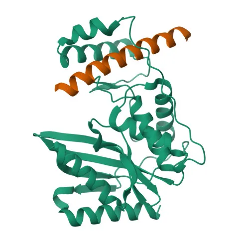
- VASH1 (vasohibin-1) is a tubulinyl-Tyr carboxypeptidase (TTCP 1) that removes the C-terminal tyrosine residue of alpha-tubulin, a key post-translational modification regulating microtubule dynamics and function. Related references: PMID:29146869 PMID:31171830 PMID:31235910
- Critical for spindle function and accurate chromosome segregation during mitosis, as microtubule detyrosination modulates mitotic spindle length and positioning. Related references: PMID:31171830
- Acts as a selective angiogenesis inhibitor: it blocks endothelial cell migration, proliferation, and tube formation without affecting smooth muscle cells or fibroblasts. Related references: PMID:15467828 PMID:16488400 PMID:16707096
- Localizes to the cytoplasm and can be secreted into the extracellular space.
- Preferentially expressed in endothelial cells; highly expressed in fetal organs, brain, and placenta, with lower levels in heart and kidney. Notably detected in microvessel endothelial cells of atherosclerotic lesions. Related references: PMID:15467828 PMID:16707096
- Subject to alternative splicing and ubiquitin-like conjugation, which may regulate its activity or stability.
Experimental Guidance and Technical Tips
- For Western blotting, consider testing a range of antibody dilutions and confirm detection of the expected ~41 kDa band in appropriate lysates (e.g., HUVEC or other endothelial cells). Validate specificity using siRNA knockdown or overexpression controls if needed.
- The polyclonal nature and immunogen spanning nearly the full-length protein (aa 1–365) suggest the antibody may recognize multiple epitopes; note that detection of post-translationally modified forms or splice variants could vary.
- Confirmed cross-reactivity with mouse allows use in mouse model systems; always validate the antibody in the relevant tissue or cell type before large-scale studies.
- For ELISA, titrate the antibody as a capture or detection reagent in your specific assay format and include appropriate positive (e.g., recombinant VASH1 protein) and negative controls.
- Pair with a suitable secondary antibody and optimize chemiluminescent or chromogenic detection conditions for best signal-to-noise ratio.
CamelBio: Your One-Stop Sourcing Bridge
CamelBio offers this anti-VASH1 polyclonal antibody as part of a comprehensive portfolio of IVD raw materials for angiogenesis, oncology, and cytoskeleton research. We support diagnostic manufacturers and laboratories with validated antibody pairs, optimized monoclonal and polyclonal antibodies, bulk ancillary reagents, and sourcing of rare targets. Whatever your assay development needs—from concept to clinic—CamelBio is your reliable one-stop partner.
REQUEST A QUOTE
Our professional team will reply to you within one business day. Please feel free to contact us!