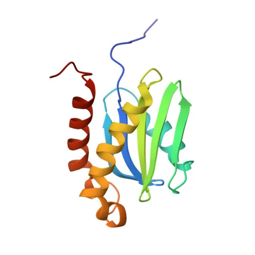
Polyclonal Antibodies
Anti-VAMP7 Polyclonal Antibody for WB, IF/ICC, ELISA - P51809
Item Number : CM0022914
Price varies based on specs and customizations
- Application
- WB, IF/ICC, ELISA
- Cross Reactivity
- Human, Mouse, Rat
- Protein Weight
- 25kDa

Shipping:
Contact us to get shipping details Enjoy On-time Dispatch Guarantee.
Why Choose Us
Easy ordering process, quality products, and dedicated support for your business success.
Core Product Specifications and Parameters
| Parameter | Value |
|---|---|
| Product Name | VAMP7 Rabbit pAb |
| Remarks/Alias | SYBL1; TIVAMP; VAMP-7; TI-VAMP; VAMP7 |
| Species | Human |
| Gene ID (Human) | 6845 |
| Gene ID | 6845 |
| Immunogen | Recombinant fusion protein containing a sequence corresponding to amino acids 11-70 of human VAMP7 (NP_005629.1) |
| Source | Rabbit |
| Category | Polyclonal Antibodies |
| Application | WB, IF/ICC, ELISA |
| Cross Reactivity | Human, Mouse, Rat |
| SWISS | P51809 |
| Protein Weight | 25kDa |
| Shipping | Ice bag |
Biological Background: VAMP7 Function and Localization
- VAMP7 (Vesicle-associated membrane protein 7), also known as synaptobrevin-like protein 1 (SYBL1) or tetanus-insensitive VAMP (TI-VAMP), is a SNARE protein involved in intracellular vesicle fusion.
- It mediates targeting and fusion of transport vesicles to target membranes during protein transport from early endosomes to lysosomes, and is required for heterotypic fusion of late endosomes with lysosomes as well as homotypic lysosomal fusion.
- VAMP7 is essential for calcium-regulated lysosomal exocytosis, contributing to processes such as chylomicron export from the ER to the Golgi, and exocytosis of mediators during eosinophil and neutrophil degranulation, and natural killer cell killing.
- The protein localizes to multiple organelles including cytoplasmic vesicles, secretory vesicle membranes, trans-Golgi network, late endosomes, lysosomes, endoplasmic reticulum, phagosome membranes, and the synapse/synaptosome.
- VAMP7 is widely expressed in all tissues tested, reflecting its fundamental role in membrane trafficking.
- Post-translational modifications include acetylation and phosphorylation, which may regulate its function and interactions in vesicle fusion events.
Experimental Guidance and Technical Tips
- The immunogen spans the N-terminal cytosolic domain (aa 11-70) of human VAMP7, which is highly conserved among human, mouse, and rat; thus, this antibody is expected to detect endogenous VAMP7 across these species in applications such as Western blotting.
- In Western blotting, a band at approximately 25 kDa corresponding to full-length VAMP7 may be observed; due to potential post-translational modifications or isoform variations, additional bands could appear—optimization of gel percentage and transfer conditions is recommended.
- For immunofluorescence or immunocytochemistry, detergent permeabilization is necessary to access the membrane-embedded antigen; co-staining with compartment-specific markers (e.g., LAMP1 for lysosomes, EEA1 for early endosomes) can confirm localization patterns.
- As a polyclonal antibody, it provides robust signal amplification but users should verify specificity with appropriate controls (e.g., siRNA knockdown or peptide blocking) in their experimental setup.
CamelBio: Your One-Stop Sourcing Bridge
CamelBio streamlines your IVD raw-material procurement by offering validated antibody pairs, optimized monoclonal and polyclonal antibodies, and bulk ancillary reagents. With expertise in rare-target sourcing, we support diagnostic manufacturers and research labs working on intracellular trafficking and lysosomal biology targets like VAMP7. From concept to clinic, CamelBio serves as your one-stop bridge for high-quality raw materials and technical services.
REQUEST A QUOTE
Our professional team will reply to you within one business day. Please feel free to contact us!