Polyclonal Antibodies
Anti-UBA6 Rabbit Polyclonal Antibody for WB, IF/ICC, IP, ELISA - A0AVT1
Item Number : CM0026263
Price varies based on specs and customizations
- Application
- WB, IF/ICC, IP, ELISA
- Cross Reactivity
- Human, Mouse
- Protein Weight
- 118kDa

Shipping:
Contact us to get shipping details Enjoy On-time Dispatch Guarantee.
Why Choose Us
Easy ordering process, quality products, and dedicated support for your business success.
Core Product Specifications and Parameters
| Parameter | Value |
|---|---|
| Product Name | UBA6 Rabbit pAb |
| Remarks/Alias | E1-L2, MOP-4, UBE1L2, UBA6 |
| Species | Human |
| GeneID | 55236 |
| Immunogen | Recombinant fusion protein containing a sequence corresponding to amino acids 1-260 of human UBA6 (NP_060697.4). |
| Source | Rabbit |
| Category | Polyclonal Antibodies |
| Application | WB, IF/ICC, IP, ELISA |
| Cross Reactivity | Human, Mouse |
| SWISS | A0AVT1 |
| Protein Weight | 118kDa |
| Shipping | Ice bag |
Biological Background: UBA6 Function and Localization
- UBA6, also known as E1-L2, MOP-4, or UBE1L2, is a ubiquitin-like modifier-activating enzyme that operates as an E1 ligase in the ubiquitin-proteasome system.
- It activates ubiquitin by adenylating its C-terminal glycine with ATP and subsequently forms a ubiquitin-E1 thioester, a critical initial step in ubiquitin conjugation. Related references: PMID:35970836 PMID:35986001
- In addition to ubiquitin, UBA6 can also activate the ubiquitin-like modifier FAT10 via adenylation of its C-terminal glycine, linking it to the UBA6 active site cysteine. Related references: PMID:17889673 PMID:35970836 PMID:35986001
- Unlike UBE1, UBA6 exhibits distinct specificity for downstream E2 enzymes; it does not charge cell cycle-associated E2s such as CDC34, indicating a specialized role in ubiquitin pathway regulation.
- UBA6 is essential for embryonic development, and its isoform 2 is predominantly expressed in testis, where it may influence spermatogenesis and male fertility.
- The protein is widely expressed across tissues and contains key functional motifs including an ATP-binding domain, metal-binding sites, and sites for acetylation and phosphorylation.
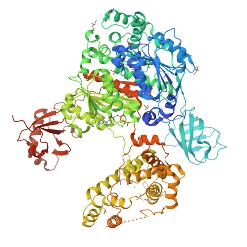
- Structurally, UBA6 has been characterized by 3D-structure determination, and it participates in the ubiquitin conjugation pathway as part of the reference proteome.
Experimental Guidance and Technical Tips
- The immunogen spans the N-terminal region (amino acids 1-260) of human UBA6, which may be critical for enzyme activity; consider this when selecting detection applications.
- For Western blot, the predicted molecular weight is 118 kDa; validate detection in lysates from human or mouse cell lines, and consider using a high-molecular-weight marker.
- For immunoprecipitation, confirm complex formation under native conditions in relevant biological samples.
- In immunofluorescence/ICC experiments, optimize permeabilization and fixation steps to detect endogenous UBA6, as localization may be influenced by cellular context.
- Although cross-reactivity is confirmed for human and mouse, it is advisable to verify reactivity in other species if needed.
CamelBio: Your One-Stop Sourcing Bridge
CamelBio serves as a one-stop procurement partner for IVD manufacturers and researchers, offering a comprehensive portfolio of raw materials including validated antibody pairs, optimized monoclonal/polyclonal antibodies, and bulk ancillary reagents. For studies focused on ubiquitin-activating enzymes like UBA6—relevant to protein degradation, cancer biology, and fertility research—CamelBio can facilitate sourcing of rare and specialized reagents to accelerate your diagnostic development.
Product Datasheet
Anti-UBA6 Rabbit Polyclonal Antibody for WB, IF/ICC, IP, ELISA - A0AVT1
REQUEST A QUOTE
Our professional team will reply to you within one business day. Please feel free to contact us!