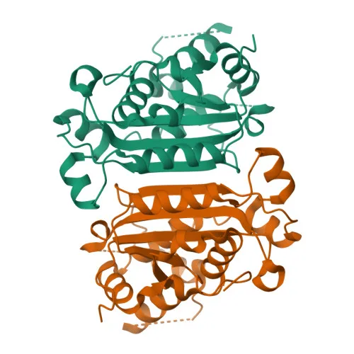
Monoclonal Antibodies
Anti-TREX1 Rabbit Monoclonal Antibody for WB, IP, ELISA - Q9NSU2
Item Number : CM0004784
Price varies based on specs and customizations
- Application
- WB, IP, ELISA
- Cross Reactivity
- Human
- Protein Weight
- 33kDa

Shipping:
Contact us to get shipping details Enjoy On-time Dispatch Guarantee.
Why Choose Us
Easy ordering process, quality products, and dedicated support for your business success.
Core Product Specifications and Parameters
| Parameter | Value |
|---|---|
| Product Name | TREX1 Rabbit mAb |
| Remarks/Alias | CRV; AGS1; DRN3; HERNS; RVCLS; TREX1 |
| Species | Human |
| GeneID (Human) | 11277 |
| Immunogen | Recombinant fusion protein containing a sequence corresponding to amino acids 150-314 of human TREX1 (Q9NSU2) |
| Source | Rabbit |
| Category | Monoclonal Antibodies |
| Application | WB, IP, ELISA |
| Cross Reactivity | Human |
| SWISS | Q9NSU2 |
| Protein Weight | 33kDa |
| Shipping | Ice bag |
Biological Background: TREX1 Function and Localization
-
TREX1, also known as 3'-5' exonuclease TREX1 or DNase III, is a major cellular 3'-to-5' DNA exonuclease that digests single-stranded DNA (ssDNA) and double-stranded DNA (dsDNA) with mismatched 3' termini. Related references: PMID:10391904 PMID:10393201 PMID:17293595
-
It prevents cell-intrinsic initiation of autoimmunity by metabolizing DNA fragments from endogenous retroelements, including L1, LTR, and SINE elements. Related references: PMID:10391904 PMID:10393201 PMID:17293595
-
TREX1 localizes to the endoplasmic reticulum membrane and is recruited to ruptured micronuclei, where it degrades DNA fragments to limit CGAS activation and subsequent type I interferon production. Related references: PMID:33476576
-
It prevents chronic ATM-dependent checkpoint activation by processing ssDNA polynucleotide species arising from aberrant DNA replication intermediates. Related references: PMID:18045533
-
Inefficiently degrades oxidized DNA, including that generated by reactive oxygen species or UV light. Related references: PMID:23993650
-
During GZMA-mediated cell death, TREX1 contributes to DNA damage in concert with NME1; NME1 nicks one strand and TREX1 removes bases from the free 3' end, enhancing damage and preventing repair. Related references: PMID:16818237
-
Subcellularly, TREX1 is found in the nucleus, cytoplasm, cytosol, and endoplasmic reticulum membrane.
-
Detected in thymus, spleen, liver, brain, heart, small intestine, and colon; mutations in TREX1 are associated with Aicardi-Goutieres syndrome, systemic lupus erythematosus, and retinal vasculopathy with cerebral leukodystrophy.
Experimental Guidance and Technical Tips
- The antibody is supplied in liquid form with ice bag shipping; aliquot and store as recommended in the datasheet upon arrival.
- For Western blot (WB), the target protein has a predicted molecular weight of 33 kDa; verify expression in human-derived samples and optimize detection conditions.
- In immunoprecipitation (IP), consider titrating the antibody concentration and include negative and positive controls to confirm specific capture of TREX1.
- For ELISA applications, optimize coating antigen and antibody dilutions to achieve the desired sensitivity and specificity.
- As cross-reactivity is validated for human only, assess reactivity in other species if required for your experimental model.
CamelBio: Your One-Stop Sourcing Bridge
CamelBio provides a comprehensive range of IVD raw materials, from validated monoclonal antibodies to bulk ancillary reagents, supporting autoimmune and genomic stability research. Our global sourcing network ensures reliable access to niche targets like TREX1, enabling seamless development of diagnostic assays. Contact our team for tailored solutions to accelerate your project from concept to clinic.
REQUEST A QUOTE
Our professional team will reply to you within one business day. Please feel free to contact us!