Polyclonal Antibodies
Anti-TP53BP2 Rabbit Polyclonal Antibody for WB, ELISA - Q13625
Item Number : CM0023463
Price varies based on specs and customizations
- Application
- WB, ELISA
- Cross Reactivity
- Human
- Protein Weight
- 126kDa

Shipping:
Contact us to get shipping details Enjoy On-time Dispatch Guarantee.
Why Choose Us
Easy ordering process, quality products, and dedicated support for your business success.
Core Product Specifications and Parameters
| Parameter | Value |
|---|---|
| Product Name | TP53BP2 Rabbit pAb |
| Remarks/Alias | BBP; 53BP2; ASPP2; P53BP2; PPP1R13A; TP53BP2 |
| Species | Human |
| GeneID (Human) | 7159 |
| GeneID | 7159 |
| Immunogen | Recombinant protein|Recombinant fusion protein containing a sequence corresponding to amino acids 200-330 of TP53BP2 (NP_001026855.2) |
| Source | Rabbit |
| Category | Polyclonal Antibodies |
| Application | WB, ELISA |
| Cross Reactivity | Human |
| SWISS | Q13625 |
| Protein Weight | 126kDa |
| Shipping | Ice bag |
Biological Background: TP53BP2 Function and Localization
- TP53BP2, also known as ASPP2, 53BP2, or Bbp, is a member of the ASPP family and undergoes alternative splicing to generate isoforms with distinct regulatory roles.
- Functions as a key regulator of apoptosis and cell growth by enhancing the DNA-binding and transactivation activity of TP53 on proapoptotic gene promoters (PubMed:12524540).
Related references:
PMID:12524540 - Inhibits the NEDD8-conjugating ability of NAE1 on CUL1, thereby decreasing NAE1-mediated apoptosis, and also impedes cell cycle progression at the G2/M phase.
- Its apoptosis-stimulating activity is inhibited by interaction with DDX42, indicating a complex regulatory network.
- Predominantly localizes to the perinuclear region of the cytoplasm and also translocates to the nucleus to exert its transcriptional co-activator function.
- Widely expressed in tissues including spleen, thymus, prostate, testis, ovary, small intestine, colon, and peripheral blood leukocytes; expression is reduced in breast carcinomas with wild-type TP53 and overexpressed in lung cancer cell lines.
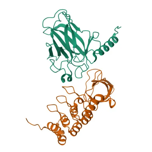
- Contains structural features including ankyrin repeats, an SH3 domain, and SH3-binding motifs, and is regulated by phosphorylation, as identified in proteomics studies.
Experimental Guidance and Technical Tips
- For Western blot, consider using whole cell lysates from human cell lines that express endogenous TP53BP2; the expected band size is approximately 126kDa.
- Validate antibody specificity in your experimental system using appropriate positive and negative controls, such as siRNA-mediated knockdown or overexpression lysates.
- Due to the polyclonal nature, it may be advisable to check lot-to-lot consistency for critical quantitative applications.
- For ELISA, optimize coating conditions and antigen concentration to achieve the best signal-to-noise ratio, especially when using recombinant protein fragments.
- Subcellular fractionation may be considered to enrich nuclear or perinuclear cytoplasmic fractions for enhanced detection of TP53BP2.
CamelBio: Your One-Stop Sourcing Bridge
This anti-TP53BP2 antibody supports apoptosis and cancer research applications involving p53 pathway regulation and cell cycle control. CamelBio serves as your comprehensive partner for IVD raw material procurement, offering validated antibody pairs, optimized monoclonal and polyclonal antibodies, bulk ancillary reagents, and rare target sourcing. Streamline your diagnostic development from concept to clinic with CamelBio's integrated supply chain and technical resources.
REQUEST A QUOTE
Our professional team will reply to you within one business day. Please feel free to contact us!