Polyclonal Antibodies
Anti-TNFRSF10A Rabbit Polyclonal Antibody for WB, IF/ICC and ELISA - O00220
Item Number : CM0016096
Price varies based on specs and customizations
- Application
- WB, IF/ICC, IF-P, ELISA
- Cross Reactivity
- Human, Mouse, Rat
- Protein Weight
- 50kDa

Shipping:
Contact us to get shipping details Enjoy On-time Dispatch Guarantee.
Why Choose Us
Easy ordering process, quality products, and dedicated support for your business success.
Core Product Specifications and Parameters
| Parameter | Value |
|---|---|
| Product Name | TNFRSF10A Rabbit pAb |
| Remarks/Alias | DR4; APO2; CD261; TRAILR1; TRAILR-1; TNFRSF10A |
| Species | Human |
| Gene ID (Human) | 8797 |
| Gene ID | 8797 |
| Immunogen | Recombinant fusion protein containing a sequence corresponding to amino acids 269-468 of human TNFRSF10A (NP_003835.3) |
| Source | Rabbit |
| Category | Polyclonal Antibodies |
| Application | WB, IF/ICC, IF-P, ELISA |
| Cross Reactivity | Human, Mouse, Rat |
| SWISS | O00220 |
| Protein Weight | 50kDa |
| Shipping | Ice bag |
Biological Background: TNFRSF10A Function and Localization
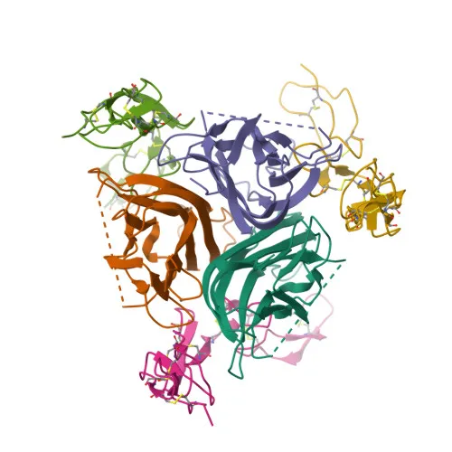
- TNFRSF10A, also known as Death Receptor 4 (DR4), TRAIL-R1, or CD261, is a member of the tumor necrosis factor receptor superfamily.
- It functions as a receptor for the cytotoxic ligand TNFSF10/TRAIL (PubMed:26457518, PubMed:38532423), triggering the formation of the death-inducing signaling complex (DISC) via the adaptor FADD, which recruits and activates caspase-8 to initiate apoptosis (PubMed:19090789). Related references: PMID:26457518 PMID:38532423 PMID:19090789
- The receptor also activates NF-kappa-B signaling, a pathway associated with cell survival and inflammation (PubMed:9430227). Related references: PMID:9430227
- TNFRSF10A is localized primarily on the cell membrane and in membrane rafts, but can also be found in the cytoplasm and cytosol.
- It is widely expressed, with high levels in spleen, peripheral blood leukocytes, small intestine, thymus, and in certain cancer cell lines such as K-562 and MCF-7.
- The protein undergoes post-translational modifications including glycosylation, disulfide bond formation, and phosphorylation, which may affect its molecular weight and function.
Experimental Guidance and Technical Tips
- The immunogen corresponds to the intracellular C-terminal region (aa 269–468) containing the death domain; therefore, for immunofluorescence applications, permeabilization of cells is typically required to access the epitope.
- In Western blotting, TNFRSF10A is predicted to migrate at ~50 kDa, but due to glycosylation, a band at a higher molecular weight may be observed; pre-incubation with PNGase F can be used to confirm glycosylation status.
- This antibody has been verified for use in WB, IF/ICC, IF-P, and ELISA, and shows cross-reactivity with mouse and rat samples, making it suitable for multi-species studies.
- For ELISA, the antibody can potentially be employed as either capture or detection antibody depending on pairing and plate format, but optimal dilutions and conditions should be determined empirically.
CamelBio: Your One-Stop Sourcing Bridge
CamelBio serves as a comprehensive sourcing platform for IVD raw materials, connecting diagnostic developers with high-quality research tools like this anti-TNFRSF10A polyclonal antibody. In addition to offering validated antibody pairs and bulk ancillary reagents, we support custom sourcing for rare targets and provide technical consulting to accelerate your assay development from concept to clinic. Explore our full portfolio of apoptosis-related reagents to find the optimal solutions for your next diagnostic project.
Product Datasheet
Anti-TNFRSF10A Rabbit Polyclonal Antibody for WB, IF/ICC and ELISA - O00220
REQUEST A QUOTE
Our professional team will reply to you within one business day. Please feel free to contact us!