Polyclonal Antibodies
Anti-TNFRSF10A Polyclonal Antibody for WB, ELISA - O00220
Item Number : CM0029628
Price varies based on specs and customizations
- Application
- WB, ELISA
- Cross Reactivity
- Human
- Protein Weight
- 50kDa

Shipping:
Contact us to get shipping details Enjoy On-time Dispatch Guarantee.
Why Choose Us
Easy ordering process, quality products, and dedicated support for your business success.
Core Product Specifications and Parameters
| Parameter | Specification |
|---|---|
| Product Name | TNFRSF10A Rabbit pAb |
| Remarks/Alias | DR4, APO2, CD261, TRAILR1, TRAILR-1, TNFRSF10A |
| Species | Human |
| GeneID (Human) | 8797 |
| GeneID | 8797 |
| Immunogen | Recombinant fusion protein containing a sequence corresponding to amino acids 269-468 of human TNFRSF10A (NP_003835.3) |
| Source | Rabbit |
| Category | Polyclonal Antibodies |
| Application | WB, ELISA |
| Cross Reactivity | Human |
| SWISS | O00220 |
| Protein Weight | 50kDa |
| Shipping | Ice bag |
Biological Background: TNFRSF10A Function and Localization
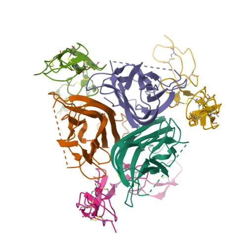
- TNFRSF10A is a tumor necrosis factor receptor superfamily member, also known as Death receptor 4 (DR4), TRAIL-R1, CD261, and APO2.
- It functions as a receptor for the cytotoxic ligand TNFSF10/TRAIL, initiating apoptosis via the extrinsic pathway. Related references: PMID:26457518, PMID:38532423
- Upon TRAIL binding, the adaptor molecule FADD recruits caspase-8 to form the death-inducing signaling complex (DISC), leading to caspase activation and programmed cell death. Related references: PMID:19090789
- TNFRSF10A also activates the NF-kappa-B signaling pathway, which can promote cell survival and inflammatory responses. Related references: PMID:9430227
- The receptor localizes predominantly at the cell membrane and within membrane rafts, with additional presence in the cytosol.
- It is widely expressed, with high levels in spleen, peripheral blood leukocytes, small intestine, thymus, and certain cancer cell lines like K-562 and MCF-7.
- Post-translational modifications include glycosylation, disulfide bond formation, phosphorylation, and methylation.
Experimental Guidance and Technical Tips
- The immunogen corresponds to amino acids 269-468 of human TNFRSF10A, encompassing part of the intracellular domain. Antibody reactivity may be specific to the cytoplasmic region; consider this when designing detection experiments.
- For WB, whole-cell lysates from TNFRSF10A-expressing cell lines such as K-562 or MCF-7 are recommended. Use appropriate negative controls (e.g., TNFRSF10A-knockout cell lines or tissues with low expression) to verify specificity.
- For ELISA, the antibody can be used for antigen capture or detection; optimize coating conditions and antibody concentrations with recombinant TNFRSF10A protein.
- Store the antibody as recommended and avoid freeze-thaw cycles. Validate performance in your specific assay system.
CamelBio: Your One-Stop Sourcing Bridge
CamelBio provides comprehensive IVD raw-material solutions, including validated antibody pairs, optimized polyclonal antibodies, and bulk ancillary reagents. For researchers investigating apoptosis and death receptor signaling, this TNFRSF10A polyclonal antibody offers reliable performance in WB and ELISA workflows. Our one-stop sourcing platform bridges the gap from discovery to clinical application, supporting you with technical consultation and supply chain expertise at every stage.
REQUEST A QUOTE
Our professional team will reply to you within one business day. Please feel free to contact us!