Polyclonal Antibodies
Anti-TNFR2/TNFRSF1B Rabbit Polyclonal Antibody for WB, ELISA - P20333
Item Number : CM0014301
Price varies based on specs and customizations
- Application
- WB, ELISA
- Cross Reactivity
- Human, Mouse, Rat
- Protein Weight
- 48kDa

Shipping:
Contact us to get shipping details Enjoy On-time Dispatch Guarantee.
Why Choose Us
Easy ordering process, quality products, and dedicated support for your business success.
Core Product Specifications and Parameters
| Parameter | Value |
|---|---|
| Product Name | TNFR2/TNFRSF1B Rabbit pAb |
| Remarks/Alias | p75; TBPII; TNFBR; TNFR2; CD120b; TNFR1B; TNFR80; TNF-R75; p75TNFR; TNF-R-II; TNFR2/TNFRSF1B |
| Species | Human |
| Gene ID (Human) | 7133 |
| Gene ID | 7133 |
| Immunogen | Synthetic peptide corresponding to amino acids 295-377 of human TNFR2/TNFRSF1B (NP_001057.1) |
| Source | Rabbit |
| Category | Polyclonal Antibodies |
| Application | WB, ELISA |
| Cross Reactivity | Human, Mouse, Rat |
| SWISS | P20333 |
| Protein Weight | 48kDa |
| Shipping | Ice bag |
Biological Background: TNFR2/TNFRSF1B Function and Localization
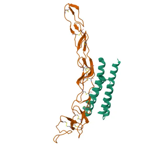
- Tumor necrosis factor receptor superfamily member 1B (TNFR2/TNFRSF1B), also known as p75, CD120b, or TNF-R-II, is encoded by the TNFRSF1B gene in humans.
- TNFR2 functions as a high-affinity receptor for tumor necrosis factor (TNF/TNFSF2) and has approximately 5-fold lower affinity for lymphotoxin-alpha (TNFSF1).
- The receptor mediates most of the metabolic effects of TNF, and its cytoplasmic domain recruits the TRAF1/TRAF2 complex, which in turn recruits the apoptotic suppressors BIRC2 and BIRC3 to modulate apoptotic signaling.
- An alternatively spliced isoform (isoform 2) acts as a dominant-negative regulator by blocking TNF-induced apoptosis, suggesting a role in fine-tuning TNF responses.
- TNFR2 is primarily localized at the cell membrane but can also be found in secreted forms, consistent with its function as a soluble decoy receptor.
- The protein undergoes several post-translational modifications, including phosphorylation and glycosylation, and contains disulfide bonds characteristic of the TNFR superfamily.
Experimental Guidance and Technical Tips
- The immunogen spans the cytoplasmic region of human TNFR2 (aa 295-377), which is conserved across species; this region may contribute to the antibody’s observed cross-reactivity with mouse and rat orthologs.
- For Western blot (WB) analysis, consider using whole-cell lysates or tissue extracts expressing TNFR2; endogenous expression may be low in some cell types, so induction or enrichment might be necessary.
- In ELISA applications, the antibody can be utilized in either direct or capture formats; however, for sandwich ELISA, a compatible antibody pair recognizing distinct epitopes should be validated.
- Given the presence of a secreted isoform, experimental design should account for both membrane-bound and soluble receptor forms when interpreting results.
CamelBio: Your One-Stop Sourcing Bridge
CamelBio supports your tumor necrosis factor receptor research and diagnostic development by offering this high-quality TNFR2/TNFRSF1B antibody along with a comprehensive portfolio of validated antibody pairs, optimized monoclonal/polyclonal antibodies, and bulk ancillary reagents. As a one-stop IVD raw-material sourcing partner, we simplify procurement from concept to clinic, including rare target raw-material sourcing to accelerate your assay development.
Product Datasheet
Anti-TNFR2/TNFRSF1B Rabbit Polyclonal Antibody for WB, ELISA - P20333
REQUEST A QUOTE
Our professional team will reply to you within one business day. Please feel free to contact us!