Monoclonal Antibodies
Anti-TNFR2/TNFRSF1B Rabbit Monoclonal Antibody for WB, ELISA - P20333
Item Number : CM0002800
Price varies based on specs and customizations
- Application
- WB, ELISA
- Cross Reactivity
- Human
- Protein Weight
- 48kDa

Shipping:
Contact us to get shipping details Enjoy On-time Dispatch Guarantee.
Why Choose Us
Easy ordering process, quality products, and dedicated support for your business success.
Core Product Specifications and Parameters
| Parameter | Value |
|---|---|
| Product Name | TNFR2/TNFRSF1B Rabbit mAb |
| Remarks/Alias | p75; TBPII; TNFBR; TNFR2; CD120b; TNFR1B; TNFR80; TNF-R75; p75TNFR; TNF-R-II; TNFR2/TNFRSF1B |
| Species | Human |
| GeneID (Human) | 7133 |
| Immunogen | Synthetic Peptide: A synthetic peptide corresponding to a sequence within amino acids 362-461 of human TNFR2/TNFRSF1B (P20333) |
| Source | Rabbit |
| Category | Monoclonal Antibodies |
| Application | WB, ELISA |
| Cross Reactivity | Human |
| SWISS | P20333 |
| Protein Weight | 48kDa |
| Shipping | Ice bag |
Biological Background: TNFR2/TNFRSF1B Function and Localization
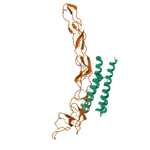
- Tumor necrosis factor receptor superfamily member 1B (TNFR2/TNFRSF1B) is a single-pass type I membrane protein, also known as p75, TNF-R-II, and CD120b.
- It functions as a high-affinity receptor for tumor necrosis factor alpha (TNF-α) and a lower-affinity receptor for homotrimeric lymphotoxin-alpha (TNFSF1).
- Upon ligand binding, the TRAF1/TRAF2 complex is recruited, which in turn recruits the apoptotic suppressors BIRC2 and BIRC3, mediating most of the metabolic effects of TNF.
- An alternative splicing isoform (Isoform 2) acts as a soluble decoy receptor that blocks TNF-induced apoptosis, suggesting a role in regulating TNF biological activity through antagonism.
- Localizes predominantly to the cell membrane, but secreted forms have also been detected.
- Known post-translational modifications include glycosylation, phosphorylation, and disulfide bond formation.
- Classified under keywords including apoptosis, alternative splicing, glycoprotein, pharmaceutical, and receptor.
Experimental Guidance and Technical Tips
- The immunogen corresponds to a peptide within amino acids 362–461 of human TNFR2, a region likely in the cytoplasmic domain. This design supports detection of both membrane-bound and intracellular forms in Western blot and ELISA.
- For Western blot, expect a band around 48 kDa under reducing conditions; validate in relevant human cell lines or tissue lysates positive for TNFR2.
- For sandwich ELISA applications, consider pairing with a validated capture antibody targeting a distinct epitope; confirm linearity and specificity using recombinant human TNFR2 or biological fluids.
CamelBio: Your One-Stop Sourcing Bridge
As a dedicated IVD raw-material partner, CamelBio supports diagnostic manufacturers and researchers from concept to clinic. Beyond this TNFR2/TNFRSF1B antibody, we offer validated antibody pairs, bulk monoclonal and polyclonal antibodies, and ancillary reagents for TNF signaling and apoptosis research. Our supply capabilities extend to rare-target raw materials, ensuring streamlined procurement for your immunological assay development.
Product Datasheet
Anti-TNFR2/TNFRSF1B Rabbit Monoclonal Antibody for WB, ELISA - P20333
REQUEST A QUOTE
Our professional team will reply to you within one business day. Please feel free to contact us!