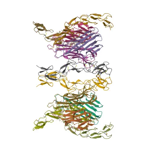
Polyclonal Antibodies
Anti-TNFR1/TNFRSF1A Polyclonal Antibody for WB, IHC-P, IF/ICC, ELISA - P19438
Item Number : CM0013310
Price varies based on specs and customizations
- Application
- WB, IHC-P, IF/ICC, ELISA
- Cross Reactivity
- Human, Mouse, Rat
- Protein Weight
- 50kDa

Shipping:
Contact us to get shipping details Enjoy On-time Dispatch Guarantee.
Why Choose Us
Easy ordering process, quality products, and dedicated support for your business success.
Core Product Specifications and Parameters
| Parameter | Value |
|---|---|
| Product Name | TNFR1/TNFRSF1A Rabbit pAb |
| Remarks/Alias | FPF; p55; p60; TBP1; TNF-R; TNFAR; TNFR1; p55-R; CD120a; TNFR55; TNFR60; TNF-R-I; TNF-R55; TNFR1/TNFRSF1A |
| Species | Human |
| GeneID (Human) | 7132 |
| GeneID | 7132 |
| Immunogen | Recombinant protein|Recombinant fusion protein containing a sequence corresponding to amino acids 345-455 of human TNFR1/TNFRSF1A (NP_001056.1). |
| Source | Rabbit |
| Category | Polyclonal Antibodies |
| Application | WB, IHC-P, IF/ICC, ELISA |
| Cross Reactivity | Human, Mouse, Rat |
| SWISS | P19438 |
| Protein Weight | 50kDa |
| Shipping | Ice bag |
Biological Background: TNFR1/TNFRSF1A Function and Localization
- TNFR1 is a member of the tumor necrosis factor receptor superfamily, also known as TNF-R1, p55, and CD120a, encoded by the TNFRSF1A gene.
- It functions as a receptor for the pro-inflammatory cytokine TNF-alpha (TNFSF2) and lymphotoxin-alpha, mediating both apoptosis and inflammatory signaling.
- Upon ligand binding, TNFR1 forms a death-inducing signaling complex (DISC) via the adaptor protein FADD, which recruits and activates caspase-8, triggering the apoptotic caspase cascade.
- TNFR1 also contributes to non-cytocidal TNF effects, including the induction of an anti-viral state and activation of acid sphingomyelinase.
- The receptor is primarily located on the cell membrane and Golgi apparatus membrane, and can be secreted as a soluble form (sTNFR1) through proteolytic cleavage.
- TNFR1 is a transmembrane glycoprotein with disulfide bonds and a signal peptide, and its gene undergoes alternative splicing.
- Mutations in TNFRSF1A are associated with periodic fever syndromes and amyloidosis, highlighting its role in inflammatory diseases.
Experimental Guidance and Technical Tips
- The immunogen corresponds to the intracellular region (aa 345-455) of human TNFR1; therefore, this antibody may detect both full-length receptor and intracellular fragments. Consider antigen retrieval for IHC to detect membrane-localized receptor.
- For Western blot, the predicted molecular weight is ~50 kDa; a band around 50 kDa is expected for full-length TNFR1. Additional bands may represent soluble forms or degradation products; validate in your sample system.
- The polyclonal antibody may cross-react with mouse and rat TNFR1 due to sequence conservation; however, verify cross-reactivity in your specific experimental setup.
- Suitable applications include WB, IHC-P, IF/ICC, and ELISA; optimize antibody concentration and blocking conditions for each application.
- For functional studies, note that TNFR1 can be shed from the cell surface; therefore, consider using appropriate controls to distinguish membrane-bound vs. soluble forms.
CamelBio: Your One-Stop Sourcing Bridge
CamelBio offers a comprehensive one-stop sourcing platform for IVD raw materials, including validated antibody pairs, optimized polyclonal and monoclonal antibodies, and bulk ancillary reagents. Our TNFR1/TNFRSF1A Rabbit pAb supports researchers studying apoptosis, inflammation, and TNF signaling, with reliable supply for diagnostic development. For rare or custom targets, our technical team provides end-to-end sourcing and consultation to accelerate your transition from concept to clinic.
Product Datasheet
Anti-TNFR1/TNFRSF1A Polyclonal Antibody for WB, IHC-P, IF/ICC, ELISA - P19438
REQUEST A QUOTE
Our professional team will reply to you within one business day. Please feel free to contact us!