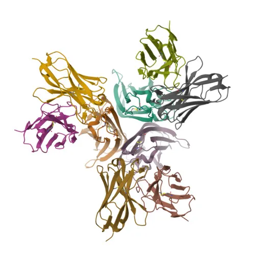
Polyclonal Antibodies
Anti-TNF-α Rabbit Polyclonal Antibody for WB, IF/ICC, IHC-P, ELISA - P01375
Item Number : CM0017885
Price varies based on specs and customizations
- Application
- WB, IF/ICC, IHC-P, ELISA
- Cross Reactivity
- Human, Mouse
- Protein Weight
- 26 kDa

Shipping:
Contact us to get shipping details Enjoy On-time Dispatch Guarantee.
Why Choose Us
Easy ordering process, quality products, and dedicated support for your business success.
Core Product Specifications and Parameters
| Parameter | Value |
|---|---|
| Product Name | TNF-α Rabbit pAb |
| Remarks/Alias | DIF; TNFA; TNFSF2; TNLG1F; TNF-alpha; TNF-α |
| Species | Human |
| GeneID (Human) | 7124 |
| GeneID | 7124 |
| Immunogen | Recombinant fusion protein containing a sequence corresponding to amino acids 134-233 of human TNF-α (NP_000585.2). |
| Source | Rabbit |
| Category | Polyclonal Antibodies |
| Application | WB, IF/ICC, IHC-P, ELISA |
| Cross Reactivity | Human, Mouse |
| SWISS | P01375 |
| Protein Weight | 26 kDa |
| Shipping | Ice bag |
Biological Background: TNF-α Function and Localization
- TNF-α is a homotrimeric cytokine that exists as both a 26 kDa type II transmembrane precursor and a 17 kDa soluble form generated by proteolytic cleavage. Its structure includes an intracellular domain, a single transmembrane helix, and an extracellular C‑terminal receptor‑binding domain.
- Encoded by the TNF gene (alias TNFA, TNFSF2) on human chromosome 6, TNF‑α is a prototypical member of the tumor necrosis factor superfamily, which comprises 19 ligands that regulate diverse biological processes.
- TNF‑α primarily signals through two receptors, TNFRSF1A/TNFR1 and TNFRSF1B/TNFBR. It is mainly secreted by activated macrophages and exerts pleiotropic effects including induction of apoptosis in certain tumor cell lines, fever (pyrogenicity), and cachexia.
- In rheumatoid arthritis, TNF‑α impairs regulatory T‑cell function by upregulating protein phosphatase 1 (PP1), which dephosphorylates the key FOXP3 residue Ser‑418, causing Treg functional disability. Related references: PMID:23396208
- TNF‑α acts as a key mediator of cell death in the anticancer action of BCG‑stimulated neutrophils combined with a DIABLO/SMAC mimetic, demonstrated in the RT4v6 bladder cancer cell line. Related references: PMID:16829952, PMID:22517918, PMID:23396208
- Synergistically with IL‑1B and IL‑6, TNF‑α induces VEGF production, thereby promoting angiogenesis. Related references: PMID:12794819
- Subcellular localization is dynamic: the full‑length protein resides at the cell membrane, while the cleaved ectodomain is secreted. The intracellular domain (ICD) can translocate to the nucleus and stimulate IL‑12 production in dendritic cells.
- Post‑translational modifications include glycosylation, myristoylation, phosphorylation, and disulfide bond formation, all of which are critical for proper folding, activity, and stability.
Experimental Guidance and Technical Tips
- The immunogen corresponds to the C‑terminal region (aa 134–233) of human TNF‑α, which lies within the mature, secreted portion. The resulting polyclonal antibody may recognize both membrane‑bound and soluble forms of the cytokine.
- For western blotting, a band at approximately 26 kDa represents the full‑length transmembrane precursor, while a band at ~17 kDa indicates the shed soluble form. Use LPS‑stimulated macrophage or monocyte lysates as positive controls.
- In immunohistochemistry, paraffin‑embedded sections may require heat‑mediated antigen retrieval. Consider testing known TNF‑α‑rich tissues such as inflamed lymph nodes or rheumatoid synovium.
- When applying immunofluorescence or ELISA, validate antibody specificity using recombinant TNF‑α or relevant positive/negative cell lines expressing the target.
CamelBio: Your One-Stop Sourcing Bridge
CamelBio provides diagnostic manufacturers, labs, and research institutes with one‑stop access to IVD raw materials, technical services, and consulting—covering every stage from concept to clinic. This TNF‑α rabbit polyclonal antibody is a versatile tool for immunoassay development in inflammation, oncology, and autoimmune disease research. Beyond antibodies, CamelBio supplies validated antigen pairs, bulk ancillary reagents, and rare‑target raw‑material sourcing, empowering you to accelerate your diagnostic assay pipeline with reliable, high‑quality critical reagents.
Product Datasheet
Anti-TNF-α Rabbit Polyclonal Antibody for WB, IF/ICC, IHC-P, ELISA - P01375
REQUEST A QUOTE
Our professional team will reply to you within one business day. Please feel free to contact us!