Polyclonal Antibodies
Anti-TNF-α Rabbit Polyclonal Antibody for WB, IF-P, IHC-P, ELISA - P01375
Item Number : CM0016171
Price varies based on specs and customizations
- Application
- WB, IF-P, IHC-P, ELISA
- Cross Reactivity
- Human, Mouse, Rat
- Protein Weight
- 26 kDa

Shipping:
Contact us to get shipping details Enjoy On-time Dispatch Guarantee.
Why Choose Us
Easy ordering process, quality products, and dedicated support for your business success.
Core Product Specifications and Parameters
| Parameter | Value |
|---|---|
| Product Name | TNF-α Rabbit pAb |
| Remarks/Alias | DIF; TNFA; TNFSF2; TNLG1F; TNF-alpha; TNF-α |
| Species | Human |
| GeneID (human) | 7124 |
| GeneID | 7124 |
| Immunogen | Synthetic peptide |
| Source | Rabbit |
| Category | Polyclonal Antibodies |
| Application | WB, IF-P, IHC-P, ELISA |
| Cross Reactivity | Human, Mouse, Rat |
| SWISS | P01375 |
| Protein Weight | 26 kDa |
| Shipping | Ice bag |
Biological Background: TNF-α Function and Localization
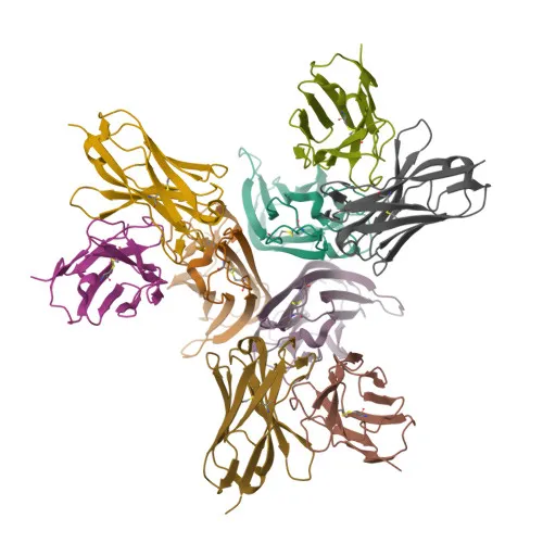
- TNF-α (cachectin, TNFSF2) is a pro-inflammatory cytokine of the tumor necrosis factor superfamily, primarily secreted by macrophages. It binds to receptors TNFRSF1A/TNFR1 and TNFRSF1B/TNFBR to mediate diverse cellular responses.
- Functions as a potent pyrogen causing fever either directly or via interleukin-1 secretion, and is implicated in the induction of cachexia. Under certain conditions, it can stimulate cell proliferation and differentiation.
- Impairs regulatory T-cell (Treg) function in rheumatoid arthritis by up-regulating protein phosphatase 1 (PP1), which dephosphorylates FOXP3 at Ser-418, thereby inactivating FOXP3. Related references: PMID:23396208
- Serves as a key mediator of cell death in the anticancer action of BCG-stimulated neutrophils combined with a DIABLO/SMAC mimetic in bladder cancer cell lines. Related references: PMID:16829952, PMID:22517918, PMID:23396208
- Plays a role in angiogenesis by synergistically inducing VEGF production with IL1B and IL6. Related references: PMID:12794819
- Induces insulin resistance in adipocytes by inhibiting insulin-induced IRS1 tyrosine phosphorylation and glucose uptake, partially through degradation of GKAP42.
- Subcellularly, TNF-α is found at the cell membrane, as a transmembrane protein, and is also secreted in a soluble form.
Experimental Guidance and Technical Tips
- The immunogen corresponds to amino acids 77-233 of human TNF (NP_000585.2), targeting a region common to both membrane-bound and soluble forms. For Western blotting, consider using stimulated macrophage lysates or recombinant protein as a positive control.
- For IHC-P, antigen retrieval optimization may be necessary; a heat-mediated citrate buffer (pH 6.0) is a suggested starting point.
- In IF-P, membrane permeabilization should be optimized to detect intracellular pools of TNF-α.
- ELISA applications may benefit from pairing this polyclonal antibody with a matched monoclonal antibody for capture-detection formats.
- Cross-reactivity with mouse and rat TNF-α is indicated, offering flexibility for translational studies, but species-specific validation is recommended.
CamelBio: Your One-Stop Sourcing Bridge
CamelBio is your one-stop sourcing partner for IVD raw materials, providing validated antibody pairs, optimized polyclonal and monoclonal antibodies, and bulk ancillary reagents to support diagnostic development from concept to clinic. For researchers focused on TNF-α and inflammatory pathways, we offer reliable access to key raw materials for assay development and validation. Let CamelBio streamline your procurement with tailored solutions for rare-target sourcing and scalable supply chains.
Product Datasheet
Anti-TNF-α Rabbit Polyclonal Antibody for WB, IF-P, IHC-P, ELISA - P01375
REQUEST A QUOTE
Our professional team will reply to you within one business day. Please feel free to contact us!