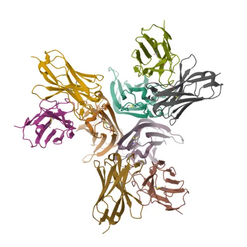
Monoclonal Antibodies
Anti-TNF-α Monoclonal Antibody for IHC-P, ELISA - P01375
Item Number : CM0007398
Price varies based on specs and customizations
- Application
- IHC-P, ELISA
- Cross Reactivity
- Human
- Protein Weight
- 26kDa

Shipping:
Contact us to get shipping details Enjoy On-time Dispatch Guarantee.
Why Choose Us
Easy ordering process, quality products, and dedicated support for your business success.
Core Product Specifications and Parameters
| Parameter | Value |
|---|---|
| Product Name | TNF-α Rabbit mAb |
| Remarks/Alias | DIF; TNFA; TNFSF2; TNLG1F; TNF-alpha; TNF-α |
| Species | Human |
| Gene ID (Human) | 7124 |
| Immunogen | Recombinant fusion protein containing a sequence corresponding to amino acids 77-233 of human TNF-α (NP_000585.2). |
| Source | Rabbit |
| Category | Monoclonal Antibodies |
| Application | IHC-P, ELISA |
| Cross Reactivity | Human |
| SWISS | P01375 |
| Protein Weight | 26kDa |
| Shipping | Ice bag |
Biological Background: TNF-α Function and Localization
- TNF-α, also known as cachectin or TNFSF2, is a member of the tumor necrosis factor (TNF) superfamily, encoded by the TNF gene in humans.
- As a key cytokine mainly secreted by macrophages, it binds to TNFRSF1A/TNFR1 and TNFRSF1B/TNFBR to mediate diverse biological effects, including cell death, inflammation, and immune regulation.
- It acts as a potent pyrogen, inducing fever, and is implicated in cachexia; under certain conditions, it can also stimulate cell proliferation and differentiation.
- TNF-α impairs regulatory T-cell (Treg) function in rheumatoid arthritis by up-regulating protein phosphatase 1 (PP1), which dephosphorylates FOXP3 at Ser-418, rendering Tregs defective (PubMed:23396208). Related references: PMID:23396208
- It promotes angiogenesis by synergistically inducing VEGF production with IL1B and IL6 (PubMed:12794819), and it drives osteoclastogenesis, linking it to bone resorption. Related references: PMID:12794819
- The TNF intracellular domain (ICD) form induces IL12 production in dendritic cells, highlighting a non-canonical signaling role.
- Subcellular localization includes the cell membrane, membrane, and secretion; it exists as a transmembrane precursor that is cleaved to release the soluble cytokine.
- Post-translational modifications, such as glycosylation, myristoylation, phosphorylation, and disulfide bond formation, are critical for its stability and function.
Experimental Guidance and Technical Tips
- The immunogen corresponds to amino acids 77-233, representing the extracellular domain of the mature, secreted form of human TNF-α. This design ensures reactivity with the soluble cytokine.
- For IHC-P, consider antigen retrieval protocols optimized for human tissue samples, and validate expression in positive controls such as inflamed lymph nodes or synovial tissue.
- In ELISA, this antibody may serve as a capture or detection reagent; establish optimal conditions by pairing with a suitable matched antibody and testing recombinant TNF-α standards.
- Given the targeted region, confirm reactivity with the native trimeric form of TNF-α in your assay system, as the immunogen covers the receptor-binding domain.
CamelBio: Your One-Stop Sourcing Bridge
CamelBio provides diagnostic manufacturers and research labs with comprehensive access to IVD raw materials, technical services, and consulting. As a trusted sourcing partner, we offer validated monoclonal antibodies like this anti-TNF-α clone, along with optimized antibody pairs, bulk ancillary reagents, and custom development for inflammation and cytokine-related biomarkers. Whether you need a specific clone for assay development or rare target raw materials, CamelBio delivers one-stop solutions from concept to clinic.
REQUEST A QUOTE
Our professional team will reply to you within one business day. Please feel free to contact us!