Polyclonal Antibodies
Anti-TMED9 Polyclonal Antibody for WB and ELISA - Q9BVK6
Item Number : CM0026699
Price varies based on specs and customizations
- Application
- WB, ELISA
- Cross Reactivity
- Human, Mouse, Rat
- Protein Weight
- 27kDa

Shipping:
Contact us to get shipping details Enjoy On-time Dispatch Guarantee.
Why Choose Us
Easy ordering process, quality products, and dedicated support for your business success.
Core Product Specifications and Parameters
| Parameter | Value |
|---|---|
| Product Name | TMED9 Rabbit pAb |
| Remarks/Alias | p25; GMP25; p24a2; HSGP25L2G; p24alpha2; TMED9 |
| Species | Human |
| Gene ID | 54732 (Human) |
| Immunogen | Recombinant fusion protein containing a sequence corresponding to amino acids 1-224 of human TMED9 (NP_059980.2) |
| Source | Rabbit |
| Category | Polyclonal Antibodies |
| Application | WB, ELISA |
| Cross Reactivity | Human, Mouse, Rat |
| SWISS | Q9BVK6 |
| Protein Weight | 27kDa |
| Shipping | Ice bag |
Biological Background: TMED9 Function and Localization
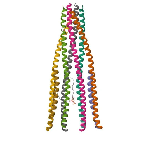
- TMED9 (Transmembrane emp24 domain-containing protein 9), also known as GMP25, p25, and p24alpha2, is a member of the p24 family of type I transmembrane proteins that cycle between the endoplasmic reticulum (ER) and the Golgi.
- It participates in vesicular protein trafficking within the early secretory pathway. In COPI vesicle-mediated retrograde transport, TMED9 contributes to coatomer recruitment to early secretory pathway membranes and enhances coatomer-dependent ARFGAP2 activity.
- TMED9 is thought to play a key role in the specific retention of p24 complexes in cis-Golgi membranes, contributing to the coupled localization of TMED2 and TMED10 in the cis-Golgi network.
- The protein may be involved in the organization of intracellular membranes, including the ER-Golgi intermediate compartment (ERGIC) and the Golgi apparatus.
- TMED9 facilitates the ER localization of the PTPN2 isoform PTPB, linking it to phosphatase trafficking.
- Subcellularly, TMED9 localizes to the endoplasmic reticulum membrane, Golgi apparatus membranes (cis- and trans-Golgi network), and the ERGIC membrane, consistent with its role in secretory pathway transport.
- Post-translational modifications include acetylation and N-linked glycosylation; the protein contains a signal peptide, a coiled-coil domain, and a single transmembrane helix.
Experimental Guidance and Technical Tips
- The immunogen corresponds to the full-length human TMED9 (residues 1–224), which encompasses the lumenal domain, transmembrane region, and cytoplasmic tail; this design may enable recognition of both native and denatured conformations.
- For Western blotting, consider that the predicted molecular weight is approximately 27 kDa, but post-translational modifications (e.g., glycosylation) could shift the observed band. Validate in relevant human, mouse, or rat cell lines expressing TMED9.
- In ELISA applications, the polyclonal nature can provide high sensitivity; however, titrations and blocking optimization are recommended for accurate quantification.
- Due to TMED9’s localization in the ER–Golgi system, consider using appropriate subcellular fractionation or immunofluorescence controls when interpreting results.
CamelBio: Your One-Stop Sourcing Bridge
CamelBio supports diagnostic manufacturers and research laboratories by providing a one-stop sourcing platform for IVD raw materials, technical services, and consulting—from concept through clinical application. With this TMED9 antibody, researchers investigating vesicular trafficking, Golgi dynamics, or secretory pathway regulation can access a proven polyclonal reagent backed by CamelBio’s comprehensive supply capabilities. Our portfolio includes validated antibody pairs, optimized monoclonal and polyclonal antibodies, bulk ancillary reagents, and rare-target raw material sourcing, all designed to streamline your assay development pipeline.
REQUEST A QUOTE
Our professional team will reply to you within one business day. Please feel free to contact us!