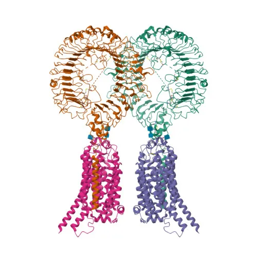
Polyclonal Antibodies
Anti-TLR7 Rabbit Polyclonal Antibody for WB, IF/ICC, IF-P, ELISA - Q9NYK1
Item Number : CM0013275
Price varies based on specs and customizations
- Application
- WB, IF/ICC, IF-P, ELISA
- Cross Reactivity
- Human, Mouse, Rat
- Protein Weight
- 121kDa

Shipping:
Contact us to get shipping details Enjoy On-time Dispatch Guarantee.
Why Choose Us
Easy ordering process, quality products, and dedicated support for your business success.
Core Product Specifications and Parameters
| Parameter | Value |
|---|---|
| Product Name | TLR7 Rabbit pAb |
| Remarks/Alias | IMD74; SLEB17; TLR7-like; TLR7 |
| Species | Human |
| GeneID (Human) | 51284 |
| GeneID | 51284 |
| Immunogen | Recombinant fusion protein containing a sequence corresponding to amino acids 30-230 of human TLR7 (NP_057646.1) |
| Source | Rabbit |
| Category | Polyclonal Antibodies |
| Application | WB, IF/ICC, IF-P, ELISA |
| Cross Reactivity | Human, Mouse, Rat |
| SWISS | Q9NYK1 |
| Protein Weight | 121kDa |
| Shipping | Ice bag |
Biological Background: TLR7 Function and Localization
- TLR7 is an endosomal receptor that recognizes uridine-containing single-stranded RNA from viruses, initiating innate and adaptive immune responses (PubMed:14976261, PubMed:32433612, PubMed:12738885).
Related references: PMID:14976261 PMID:32433612 PMID:12738885 - Ligand binding induces TLR7 dimerization and recruitment of the MYD88 adapter, forming the Myddosome complex and leading to NF-kB and IRF7 activation and subsequent cytokine/interferon production (PubMed:27742543, PubMed:32706371).
Related references: PMID:27742543 PMID:32706371 - TLR7 localizes to the endoplasmic reticulum, endosomes, lysosomes, and phagosomes, positioning it for detection of internalized pathogens.
- It is highly expressed in plasmacytoid dendritic cells, peripheral blood mononuclear cells, and tissues such as brain, placenta, spleen, and lung (PubMed:32706371).
Related references: PMID:32706371 - As a member of the Toll-like receptor family, TLR7 contains an extracellular leucine-rich repeat domain for ligand binding and a cytoplasmic TIR domain for signaling.
- Post-translational modifications and genetic variants of TLR7 are implicated in systemic lupus erythematosus, underlining its role in autoimmunity.
Experimental Guidance and Technical Tips
- The immunogen corresponds to the extracellular domain (amino acids 30–230), making this antibody suitable for recognizing full-length TLR7; consider using endosome-enriched lysates for optimal detection.
- For Western blot, run reducing SDS-PAGE and probe for a band at approximately 121 kDa; validate the signal in TLR7-expressing cell lines or tissues.
- For immunofluorescence and IF-P, include a permeabilization step (e.g., 0.1% Triton X-100) to access intracellular compartments; co-stain with organelle markers to confirm endosomal/lysosomal localization.
- In ELISA applications, test the antibody as both capture and detection reagent; pair with a matched detection antibody and titrate with recombinant TLR7 standard.
- When using with mouse or rat samples, verify reactivity empirically due to inter-species sequence differences; run a positive control alongside the experimental samples.
CamelBio: Your One-Stop Sourcing Bridge
CamelBio specializes in providing one-stop IVD raw material solutions for researchers and manufacturers developing assays in the innate immunity and infectious disease fields. Our catalog includes validated anti-TLR7 antibodies, matched antibody pairs, and bulk ancillary reagents to streamline your diagnostic development. For unique or high-demand targets, our team can source specialized materials and offer technical consulting from initial research to clinical validation.
Product Datasheet
Anti-TLR7 Rabbit Polyclonal Antibody for WB, IF/ICC, IF-P, ELISA - Q9NYK1
REQUEST A QUOTE
Our professional team will reply to you within one business day. Please feel free to contact us!