Polyclonal Antibodies
Anti-TLR1 Polyclonal Antibody for WB, IHC-P, IF/ICC, ELISA - Q15399
Item Number : CM0015943
Price varies based on specs and customizations
- Application
- WB, IHC-P, IF/ICC, ELISA
- Cross Reactivity
- Human, Mouse, Rat
- Protein Weight
- 90kDa

Shipping:
Contact us to get shipping details Enjoy On-time Dispatch Guarantee.
Why Choose Us
Easy ordering process, quality products, and dedicated support for your business success.
Core Product Specifications and Parameters
| Parameter | Value |
|---|---|
| Product Name | TLR1 Rabbit pAb |
| Remarks/Alias | TIL; CD281; rsc786; TIL. LPRS5; TLR1 |
| Species | Human |
| Gene ID (Human) | 7096 |
| Immunogen | Recombinant fusion protein containing a sequence corresponding to amino acids 30-290 of human TLR1 (NP_003254.2). |
| Source | Rabbit |
| Category | Polyclonal Antibodies |
| Application | WB, IHC-P, IF/ICC, ELISA |
| Cross Reactivity | Human, Mouse, Rat |
| SWISS | Q15399 |
| Protein Weight | 90kDa |
| Shipping | Ice bag |
Biological Background: TLR1 Function and Localization
- Encodes Toll-like receptor 1 (TLR1), also known as CD281 or TIL, a member of the Toll-like receptor family that plays a critical role in pathogen recognition and activation of innate immunity.
- Cooperates with TLR2 to recognize bacterial lipoproteins; specifically, the TLR2:TLR1:CD14 activation cluster detects triacylated lipopeptides, initiating downstream signaling. Related references: PMID:21078852 PMID:16880211
- Signaling occurs through the adaptor MYD88 and TRAF6, leading to NF-kappa-B activation and the secretion of pro-inflammatory cytokines.
- Localizes to the cell membrane and is also found in cytoplasmic vesicles, phagosome membranes, membrane rafts, and the Golgi apparatus, facilitating immune surveillance.
- Ubiquitously expressed with highest levels in spleen, ovary, peripheral blood leukocytes, thymus, and small intestine.
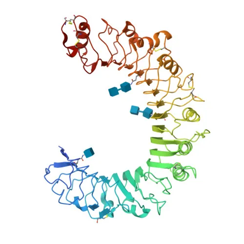
- Contains leucine-rich repeats, a transmembrane domain, and a TIR domain; post-translational modifications include glycosylation and disulfide bond formation.
Experimental Guidance and Technical Tips
- The immunogen corresponds to the extracellular domain (amino acids 30-290), suggesting the antibody may detect both the mature and precursor forms.
- In Western blotting, a band around 90 kDa is expected; due to glycosylation, the observed molecular weight may appear higher.
- For IHC-P, consider optimizing antigen retrieval techniques to achieve robust staining.
- In IF/ICC, expect membrane and vesicular-like staining patterns consistent with reported subcellular localization.
- Cross-reactivity with mouse and rat is indicated, but validation in the relevant species is recommended.
CamelBio: Your One-Stop Sourcing Bridge
CamelBio provides diagnostic manufacturers and research institutions with one-stop access to IVD raw materials, including validated antibody pairs, optimized polyclonal antibodies like this anti-TLR1 antibody, bulk ancillary reagents, and rare target sourcing. Our expertise spans innate immunity and inflammatory response research, supporting your projects from concept to clinic.
REQUEST A QUOTE
Our professional team will reply to you within one business day. Please feel free to contact us!