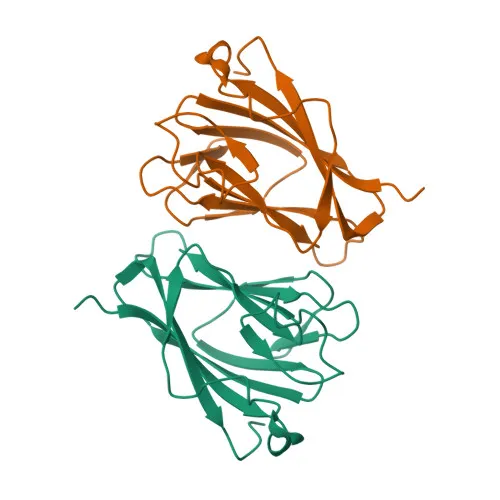
Monoclonal Antibodies
Anti-TIFA Monoclonal Antibody for WB, IHC-P, ELISA - Q793I8
Item Number : CM0011976
Price varies based on specs and customizations
- Application
- WB, IHC-P, ELISA
- Cross Reactivity
- Mouse
- Protein Weight
- 21 kDa

Shipping:
Contact us to get shipping details Enjoy On-time Dispatch Guarantee.
Why Choose Us
Easy ordering process, quality products, and dedicated support for your business success.
Core Product Specifications and Parameters
| Parameter | Value |
|---|---|
| Product Name | [KO Validated] TIFA Rabbit mAb |
| Remarks/Alias | T2bp |
| Species | Mouse |
| GeneID (Human) | 92610 |
| GeneID (Mouse) | 211550 |
| Immunogen | Synthetic peptide|A synthetic peptide corresponding to a sequence within amino acids 1-184 of mouse TIFA (NP_001343359.1). |
| Source | Rabbit |
| Category | Monoclonal Antibodies |
| Application | WB, IHC-P, ELISA |
| Cross Reactivity | Mouse |
| SWISS | Q793I8 |
| Protein Weight | 21 kDa |
| Shipping | Ice bag |
Biological Background: TIFA Function and Localization
- TIFA (TRAF-interacting protein with FHA domain-containing protein A), also known as TRAF2-binding protein, is an adapter molecule that activates pro-inflammatory NF-κB signaling after bacterial pathogen-associated molecular pattern (PAMP) detection (PubMed:11798190). Related references: PMID:11798190
- It induces TRAF6 oligomerization and polyubiquitination, leading to TAK1 and IKK activation through a proteasome-independent mechanism.
- TIFA-dependent innate immune response is specifically triggered by the bacterial metabolite ADP-D-glycero-β-D-manno-heptose (ADP-Heptose), which is recognized by ALPK1, resulting in TIFA phosphorylation at Thr-9 and homooligomerization.
- Subcellular localization is predominantly cytoplasmic, consistent with its role as a signaling adaptor.
- Post-translational modifications include phosphorylation at Thr-9 by ALPK1, a critical event for TIFA activation and downstream NF-κB signaling (By similarity).
- Tissue expression analysis shows high TIFA levels in spleen, with lower expression in heart, brain, lung, liver, kidney, and testes.
- UniProt keywords: 3D-structure, Cytoplasm, Immunity, Innate immunity, Phosphoprotein, Reference proteome.
Experimental Guidance and Technical Tips
- The immunogen corresponds to a peptide within residues 1–184 of mouse TIFA; the antibody may recognize both full-length and truncated forms if present. For WB, expect a band around 21 kDa.
- Due to KO validation, this antibody offers enhanced specificity for detecting endogenous TIFA in mouse samples; consider using knockout controls for absolute target confirmation.
- For IHC-P, validate antigen retrieval conditions in the relevant tissue; spleen is a well-suited positive control given high expression.
- In ELISA applications, coat the plate with appropriately diluted immunogen or recombinant protein, and titrate the antibody to optimize signal-to-noise ratio.
- When studying TIFA phosphorylation at Thr-9, note that activation by ADP-Heptose occurs quickly; include appropriate bacterial stimulation conditions in your experimental design.
CamelBio: Your One-Stop Sourcing Bridge
CamelBio delivers a comprehensive portfolio of IVD raw materials spanning innate immunity, cell signaling, and infectious disease targets — including this validated anti-TIFA monoclonal antibody. We support diagnostic manufacturers with one‑stop access to bulk quantities, customized antibody development, and technical solutions to accelerate your assay development. Contact us to discuss how our sourcing capabilities can meet your TIFA‑related and broader NF‑κB pathway reagent needs efficiently.
REQUEST A QUOTE
Our professional team will reply to you within one business day. Please feel free to contact us!