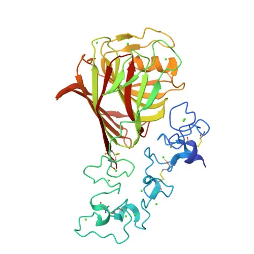
Monoclonal Antibodies
Anti-THBS1 Rabbit Monoclonal Antibody for WB - P07996
Item Number : CM0010494
Price varies based on specs and customizations
- Application
- WB, IP, IHC-P, ELISA
- Cross Reactivity
- Human, Mouse, Rat
- Protein Weight
- 129 kDa/120 kDa

Shipping:
Contact us to get shipping details Enjoy On-time Dispatch Guarantee.
Why Choose Us
Easy ordering process, quality products, and dedicated support for your business success.
Core Product Specifications and Parameters
| Parameter | Value |
|---|---|
| Product Name | THBS1 Rabbit mAb |
| Remarks/Alias | TSP; THBS; TSP1; TSP-1; THBS-1 |
| Species | Human |
| GeneID (Human) | 7057 |
| GeneID | 7057 |
| Immunogen | A synthetic peptide corresponding to amino acids 19-34 of human THBS1 (NP_003237.2) |
| Source | Rabbit |
| Category | Monoclonal Antibodies |
| Application | WB, IP, IHC-P, ELISA |
| Cross Reactivity | Human, Mouse, Rat |
| SWISS | P07996 |
| Protein Weight | 129 kDa/120 kDa |
| Shipping | Ice bag |
Biological Background: Thrombospondin-1 Function and Localization
- THBS1, also known as Glycoprotein G, is an adhesive glycoprotein that mediates cell-to-cell and cell-to-matrix interactions.
Related references: PMID:15014436 PMID:18285447 PMID:2430973 - Multifunctional protein involved in inflammation, angiogenesis, wound healing, ROS signaling, NO signaling, apoptosis, senescence, aging, stem cell self-renewal, and metabolic homeostasis.
Related references: PMID:10613822 PMID:11134179 PMID:1371676 - Negatively modulates dendritic cell activation and cytokine release, contributing to the resolution of inflammation and immune homeostasis.
Related references: PMID:14568985 - Ligand for receptors CD47, CD36, and SIRPA, mediating effects on NO signaling, ROS production, cellular senescence, apoptosis, and angiogenesis suppression.
Related references: PMID:19004835 PMID:8550562 PMID:29042481 PMID:10613822 PMID:11134179 PMID:1371676 PMID:24511121 - May play a role in metabolic dysfunction in diet-induced obesity and ER stress response via interactions with ATF6.
- Subcellular localization includes secretion, cell surface, extracellular matrix, endoplasmic reticulum, and sarcoplasmic reticulum.
- Tissue specificity: Expressed by platelets (at protein level) and by monocyte-derived immature and mature dendritic cells (at protein level).
Related references: PMID:101549 PMID:14568985 - Keywords: Angiogenesis, Apoptosis, Calcium, Cell adhesion, Disulfide bond, EGF-like domain, Endoplasmic reticulum, Extracellular matrix, Glycoprotein, Heparin-binding, Inflammatory response, Secreted, Signal, Unfolded protein response.
Experimental Guidance and Technical Tips
- The immunogen corresponds to amino acids 19-34 of human THBS1. For Western blot, consider using reducing conditions, as THBS1 contains disulfide bonds; the observed band may appear at approximately 120 kDa (reducing) or at the full-length 129 kDa.
- Validate the antibody in relevant human, mouse, or rat samples, such as platelet lysates or dendritic cell extracts.
- For immunohistochemistry on paraffin-embedded sections, antigen retrieval may be necessary to expose the epitope.
- Optimize antibody dilution for IP and ELISA by titration in your experimental system.
CamelBio: Your One-Stop Sourcing Bridge
CamelBio provides diagnostic manufacturers, labs, and research institutes with one-stop access to IVD raw materials, technical services, and consulting—covering every stage from concept to clinic. This anti-THBS1 monoclonal antibody supports research into inflammation, angiogenesis, and metabolic disorders. CamelBio offers validated antibody pairs, optimized monoclonal/polyclonal antibodies, bulk ancillary reagents, and rare target raw-material sourcing to streamline your assay development.
REQUEST A QUOTE
Our professional team will reply to you within one business day. Please feel free to contact us!