Monoclonal Antibodies
Anti-TGFBI (TGF beta induced) Rabbit Monoclonal Antibody for WB, ELISA - Q15582
Item Number : CM0004300
Price varies based on specs and customizations
- Application
- WB, ELISA
- Cross Reactivity
- Human, Mouse, Rat
- Protein Weight
- 75 kDa

Shipping:
Contact us to get shipping details Enjoy On-time Dispatch Guarantee.
Why Choose Us
Easy ordering process, quality products, and dedicated support for your business success.
Core Product Specifications and Parameters
| Parameter | Value |
|---|---|
| Product Name | TGF beta induced (TGFBI) Rabbit mAb |
| Remarks/Alias | CSD; CDB1; CDG2; CSD1; CSD2; CSD3; EBMD; LCD1; BIGH3; CDGG1; TGF beta induced (TGFBI) |
| Species | Human |
| Gene ID (Human) | 7045 |
| Gene ID | 7045 |
| Immunogen | A synthetic peptide corresponding to amino acids 550-650 of human TGF beta induced (TGFBI) (NP_000349.1) |
| Source | Rabbit |
| Category | Monoclonal Antibodies |
| Application | WB, ELISA |
| Cross Reactivity | Human, Mouse, Rat |
| SWISS | Q15582 |
| Protein Weight | 75 kDa |
| Shipping | Ice bag |
Biological Background: TGFBI Function and Localization
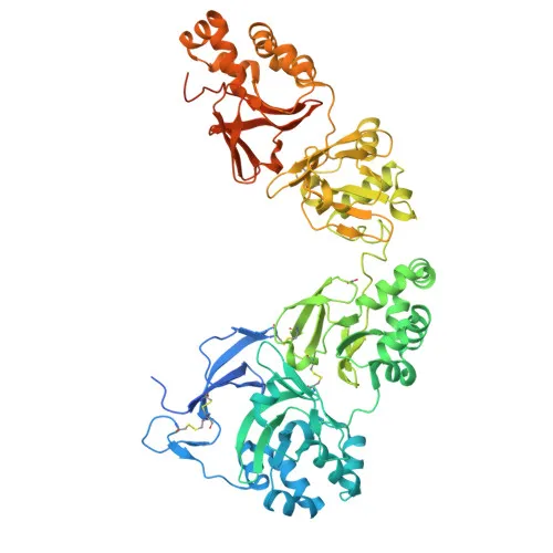
- Protein identity: TGFBI (transforming growth factor-beta-induced protein ig-h3), also known as kerato-epithelin and RGD-CAP (RGD-containing collagen-associated protein), is a secreted extracellular matrix protein encoded by the TGFBI gene (also known as BIGH3).
- Functions in cell adhesion and collagen interactions: TGFBI mediates cell adhesion and may play a role in cell-collagen interactions, contributing to extracellular matrix organization. Related references: PMID:8024701
- Subcellular localization: The protein is secreted into the extracellular space and associates with the extracellular matrix.
- Tissue-specific expression: TGFBI is highly expressed in the corneal epithelium, with lower expression detected in heart, placenta, lung, liver, skeletal muscle, kidney, and pancreas. Related references: PMID:27609313 PMID:8077289
- Disease relevance: Mutations in TGFBI are linked to various corneal dystrophies, and the protein can form amyloid deposits in affected tissues.
- Structural and post-translational features: TGFBI contains an RGD motif that facilitates integrin binding. Key post-translational modifications include gamma-carboxylation and phosphorylation, and it contains disulfide bonds.
- Additional keywords: Cell adhesion, extracellular matrix, secreted, amyloid, amyloidosis, corneal dystrophy, sensory transduction, vision.
Experimental Guidance and Technical Tips
- The immunogen corresponds to amino acids 550–650 of human TGFBI (C‑terminal region); the monoclonal antibody likely detects an epitope within this region. Validate detection in the species and sample system of interest (human, mouse, or rat).
- For Western blot (WB), consider using whole‑cell lysates or tissue extracts known to express TGFBI (e.g., corneal epithelium, placenta). The predicted protein weight is approximately 75 kDa.
- For ELISA, the antibody may be used as a capture or detection reagent; optimization of coating concentration and detection conditions is recommended.
- Because TGFBI is a secreted ECM protein, evaluate both cell‑associated and soluble fractions as needed, depending on the experimental context.
CamelBio: Your One-Stop Sourcing Bridge
CamelBio supports diagnostic manufacturers and laboratories with a comprehensive portfolio of IVD raw materials, including validated antibody pairs, optimized monoclonal and polyclonal antibodies, and bulk ancillary reagents. For targets like TGFBI, central to corneal dystrophy and extracellular matrix research, we provide one‑stop sourcing—from concept to clinic—along with technical consulting and supply of rare‑target raw materials.
Product Datasheet
Anti-TGFBI (TGF beta induced) Rabbit Monoclonal Antibody for WB, ELISA - Q15582
REQUEST A QUOTE
Our professional team will reply to you within one business day. Please feel free to contact us!