Polyclonal Antibodies
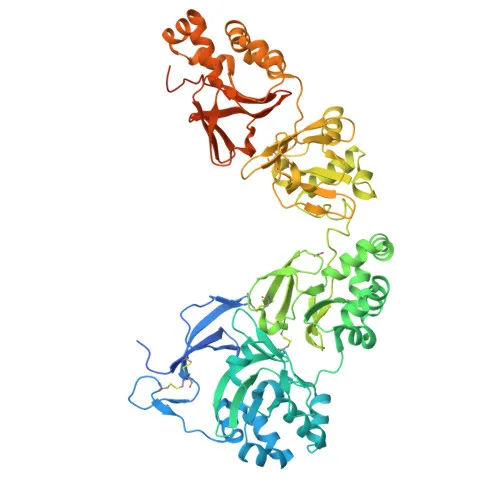
Anti-TGFBI Polyclonal Antibody for WB, IHC-P, IF/ICC, ELISA - Q15582
Item Number : CM0029759
Price varies based on specs and customizations
- Application
- WB, IHC-P, IF/ICC, ELISA
- Cross Reactivity
- Human, Mouse, Rat
- Protein Weight
- 75kDa

Shipping:
Contact us to get shipping details Enjoy On-time Dispatch Guarantee.
Why Choose Us
Easy ordering process, quality products, and dedicated support for your business success.
Core Product Specifications and Parameters
| Parameter | Description |
|---|---|
| Product Name | TGF beta induced (TGFBI) Rabbit pAb |
| Remarks/Alias | CSD; CDB1; CDG2; CSD1; CSD2; CSD3; EBMD; LCD1; BIGH3; CDGG1; TGF beta induced (TGFBI) |
| Species | Human |
| Gene ID (Human) | 7045 |
| Gene ID | 7045 |
| Immunogen | Synthetic peptide corresponding to a sequence within amino acids 550-650 of human TGF beta induced (TGFBI) (NP_000349.1). |
| Source | Rabbit |
| Category | Polyclonal Antibodies |
| Application | WB, IHC-P, IF/ICC, ELISA |
| Cross Reactivity | Human, Mouse, Rat |
| SWISS | Q15582 |
| Protein Weight | 75kDa |
| Shipping | Ice bag |
Biological Background: TGFBI Function and Localization
- TGFBI (transforming growth factor-beta-induced) encodes a secreted protein known as kerato-epithelin, RGD-containing collagen-associated protein (RGD-CAP), and BIGH3.
- It functions in cell adhesion, mediated through RGD motifs, as demonstrated by experimental evidence (PubMed:8024701). Related references: PMID:8024701
- The protein is suggested to be involved in cell-collagen interactions (By similarity).
- TGFBI localizes to the extracellular space and extracellular matrix upon secretion.
- It exhibits high expression in the corneal epithelium, with additional expression in heart, placenta, lung, liver, skeletal muscle, kidney, and pancreas.
- Post-translational modifications include phosphorylation and gamma-carboxyglutamic acid formation, and the protein contains disulfide bonds.
- Disease-associated mutations are linked to various corneal dystrophies and amyloidosis, underscoring its role in vision and sensory transduction.
Experimental Guidance and Technical Tips
- The immunogen was designed using a synthetic peptide covering amino acids 550–650 of human TGFBI. This segment can be employed in peptide blocking experiments to confirm signal specificity or to map the epitope recognized by the antibody.
- For Western blot analysis, consider using cell lines or tissues with known high TGFBI expression, such as corneal epithelial cells, to optimize detection conditions. The predicted molecular weight from the immunogen region is approximately 75 kDa.
- In IHC-P and IF/ICC applications, validate staining patterns in positive control tissues like cornea; include isotype controls and consider antigen retrieval methods to enhance sensitivity.
- For ELISA, the peptide immunogen may serve as a standard for quantitative assays. Titrate the antibody and optimize coating conditions for linear detection range.
CamelBio: Your One-Stop Sourcing Bridge
CamelBio offers diagnostic manufacturers and laboratories streamlined access to premium IVD raw materials, including this anti-TGFBI polyclonal antibody for research in cell adhesion, extracellular matrix biology, and corneal dystrophy. Our portfolio features validated antibody pairs, high-specificity monoclonal and polyclonal antibodies, and bulk ancillary reagents, bolstered by specialized sourcing for rare-target antibodies to support end-to-end assay development.
REQUEST A QUOTE
Our professional team will reply to you within one business day. Please feel free to contact us!