Monoclonal Antibodies
Anti-TGFBI (BIGH3) Monoclonal Antibody for WB, IHC-P, ELISA - Q15582
Item Number : CM0006405
Price varies based on specs and customizations
- Application
- WB, IHC-P, ELISA
- Cross Reactivity
- Human, Mouse, Rat
- Protein Weight
- 74KDa

Shipping:
Contact us to get shipping details Enjoy On-time Dispatch Guarantee.
Why Choose Us
Easy ordering process, quality products, and dedicated support for your business success.
Core Product Specifications and Parameters
| Parameter | Value |
|---|---|
| Product Name | TGF beta induced (TGFBI) Rabbit mAb |
| Remarks/Alias | CSD; CDB1; CDG2; CSD1; CSD2; CSD3; EBMD; LCD1; BIGH3; CDGG1; TGF beta induced (TGFBI) |
| Species | Human |
| GeneID (human) | 7045 |
| GeneID | 7045 |
| Immunogen | A synthetic peptide corresponding to a sequence within amino acids 550-650 of human TGFBI (NP_000349.1) |
| Source | Rabbit |
| Category | Monoclonal Antibodies |
| Application | WB, IHC-P, ELISA |
| Cross Reactivity | Human, Mouse, Rat |
| SWISS | Q15582 |
| Protein Weight | 74KDa |
| Shipping | Ice bag |
Biological Background: TGFBI Function and Localization
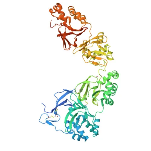
- TGFBI (transforming growth factor-beta-induced protein ig-h3), also known as kerato-epithelin, BIGH3, and RGD-CAP, is a secreted extracellular matrix protein.
- It plays a role in cell adhesion, mediating cell attachment through interaction with integrins (PubMed:8024701). Related references: PMID:8024701
- May contribute to cell-collagen interactions, suggesting involvement in matrix organization and tissue integrity.
- Localized to the extracellular space and extracellular matrix, consistent with its function as a matricellular protein.
- Highly expressed in the corneal epithelium, and also found in heart, placenta, lung, liver, skeletal muscle, kidney, and pancreas, indicating a broad tissue distribution.
- Mutations in TGFBI are associated with corneal dystrophies (e.g., granular, lattice, and Avellino types), and the protein can form amyloid deposits in certain pathological conditions.
- Post-translational modifications include gamma-carboxyglutamic acid, phosphorylation, and disulfide bonds, which may regulate its structure and interactions.
Experimental Guidance and Technical Tips
- The antibody was raised against a synthetic peptide spanning amino acids 550–650, a region potentially conserved across human, mouse, and rat, which may account for the observed cross-reactivity.
- For Western blot, consider running reduced and non-reduced samples to evaluate the impact of predicted disulfide bonds on electrophoretic mobility.
- When performing IHC-P, optimize antigen retrieval conditions (e.g., heat-induced epitope retrieval with citrate or EDTA buffer) and titrate the primary antibody concentration based on the tissue type and fixation method.
- In ELISA, include appropriate controls to confirm specificity, particularly if analyzing complex biological fluids.
CamelBio: Your One-Stop Sourcing Bridge
CamelBio supports diagnostic manufacturers and research laboratories with comprehensive sourcing of IVD raw materials for cell adhesion and corneal dystrophy applications. Our portfolio features validated antibody pairs, monoclonal and polyclonal antibodies, and bulk ancillary reagents to accelerate assay development. For challenging targets like TGFBI, we offer customized raw-material procurement services, bridging the gap from concept to clinic.
REQUEST A QUOTE
Our professional team will reply to you within one business day. Please feel free to contact us!