Polyclonal Antibodies
Anti-TGF beta Receptor II (TGFBR2) Rabbit Polyclonal Antibody for WB, IHC-P, IF/ICC, ELISA - P37173
Item Number : CM0014594
Price varies based on specs and customizations
- Application
- WB, IHC-P, IF/ICC, ELISA
- Cross Reactivity
- Human, Mouse, Rat
- Protein Weight
- 65kDa

Shipping:
Contact us to get shipping details Enjoy On-time Dispatch Guarantee.
Why Choose Us
Easy ordering process, quality products, and dedicated support for your business success.
Core Product Specifications and Parameters
| Parameter | Value |
|---|---|
| Product Name | TGF beta Receptor II Rabbit pAb |
| Remarks/Alias | AAT3; FAA3; LDS2; MFS2; RIIC; LDS1B; LDS2B; TAAD2; TBRII; TBR-ii; TGFR-2; tbetaR-II; TGFbeta-RII; TGF beta Receptor II (TGFBR2) |
| Species | Human |
| GeneID (human) | 7048 |
| GeneID | 7048 |
| Immunogen | Recombinant protein|Recombinant fusion protein containing a sequence corresponding to amino acids 23-280 of human TGF beta Receptor II (TGFBR2) (NP_003233.4). |
| Source | Rabbit |
| Category | Polyclonal Antibodies |
| Application | WB, IHC-P, IF/ICC, ELISA |
| Cross Reactivity | Human, Mouse, Rat |
| SWISS | P37173 |
| Protein Weight | 65kDa |
| Shipping | Ice bag |
Biological Background: TGF-beta Receptor Type-2 Function and Localization
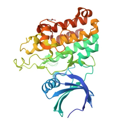
- Receptor identity and structure: TGF-beta receptor type-2 (TGFBR2) is a single-pass type I transmembrane serine/threonine kinase that serves as the high-affinity receptor for TGF-beta cytokines TGFB1, TGFB2, and TGFB3. It is encoded by the TGFBR2 gene on human chromosome 3, with alternative splicing producing multiple isoforms, some of which are linked to hereditary nonpolyposis colorectal cancer, aortic aneurysm, and craniosynostosis.
- Canonical SMAD-dependent signaling: Upon ligand binding, TGFBR2 pairs with two type I receptors (TGFBR1) to form a heterotetrameric complex. The constitutively active kinase domain of TGFBR2 phosphorylates TGFBR1, which in turn activates SMAD2 and SMAD3. The phosphorylated SMADs complex with SMAD4 and translocate to the nucleus to regulate TGF-beta target gene transcription, controlling cell cycle arrest, apoptosis, differentiation, and extracellular matrix production.
- Non-canonical signaling: In addition to the canonical SMAD pathway, TGFBR2 engages SMAD-independent cascades, including MAP kinase, PI3K/AKT, and Rho-like GTPase pathways, to modulate cell migration, proliferation, and survival.
- Ligand binding and affinity: TGFBR2 binds TGFB1, TGFB2, and TGFB3 in the picomolar affinity range without requiring co-receptors, mediating the initial step of TGF-beta signal transduction.
- Subcellular localization: TGFBR2 primarily localizes to the cell membrane and membrane rafts, with evidence of a secreted form detected in extracellular environments, consistent with its role in cell-to-cell communication.
- Post-translational modifications: The receptor is a glycoprotein with multiple intramolecular disulfide bonds essential for its tertiary structure. It undergoes phosphorylation at serine and threonine residues within its kinase domain and cytoplasmic tail, which modulates activity and downstream signaling.
- Keywords summary: The UniProt record highlights its involvement in apoptosis, growth regulation, differentiation, and disease associations including aortic aneurysm, craniosynostosis, and hereditary nonpolyposis colorectal cancer.
Experimental Guidance and Technical Tips
- Immunogen design: The antibody was raised against a recombinant protein encompassing amino acids 23–280 of human TGFBR2, which covers the N-terminal extracellular ligand-binding domain. This region is relatively conserved across human, mouse, and rat, explaining the observed cross-reactivity; however, endogenous glycosylation patterns may influence epitope accessibility in native samples.
- Western blot (WB) considerations: The predicted molecular weight of TGFBR2 is approximately 65 kDa; however, due to glycosylation, you may observe a band shift. Use reducing conditions for clear resolution. Validate in positive control lysates (e.g., HeLa, A549) known to express TGFBR2.
- Immunohistochemistry (IHC-P) tips: For paraffin-embedded sections, optimize antigen retrieval (e.g., citrate buffer pH 6.0 or EDTA pH 9.0) and consider testing multiple concentrations of primary antibody (typical range 1:50–1:500). Membrane staining is expected; inclusion of a phosphatase inhibitor in blocking buffers may help preserve phosphorylation-dependent epitopes.
- Immunofluorescence (IF/ICC) guidance: Because TGFBR2 is a transmembrane receptor, permeabilization with Triton X-100 or saponin is necessary for intracellular epitope detection. Co-staining with membrane or organelle markers is recommended to confirm localization. The antibody may also recognize the secreted form, so careful fixation is advised to avoid extracellular background.
- ELISA usage: This polyclonal antibody can be explored as a capture or detection reagent in sandwich ELISA formats. Perform checkerboard titrations to determine optimal coating (1–10 µg/mL) and detection concentrations, and validate with recombinant TGFBR2 protein standards.
CamelBio: Your One-Stop Sourcing Bridge
CamelBio offers a streamlined, single-vendor experience for diagnostic manufacturers and research laboratories seeking high-quality raw materials for TGF-beta signaling studies, fibrosis, and oncology assay development. We supply validated antibody pairs, optimized monoclonal and polyclonal antibodies like this TGFBR2 rabbit pAb, bulk ancillary reagents, and custom raw-material sourcing. Whether you are scaling up an existing immunoassay or developing a novel biomarker detection panel, CamelBio serves as your integrated partner from concept to clinic.
Product Datasheet
Anti-TGF beta Receptor II (TGFBR2) Rabbit Polyclonal Antibody for WB, IHC-P, IF/ICC, ELISA - P37173
REQUEST A QUOTE
Our professional team will reply to you within one business day. Please feel free to contact us!