Monoclonal Antibodies
Anti-TGF beta Receptor II Monoclonal Antibody for WB - P37173
Item Number : CM0003567
Price varies based on specs and customizations
- Application
- WB, ELISA
- Cross Reactivity
- Human
- Protein Weight
- 65kDa/67kDa/9kDa

Shipping:
Contact us to get shipping details Enjoy On-time Dispatch Guarantee.
Why Choose Us
Easy ordering process, quality products, and dedicated support for your business success.
Core Product Specifications and Parameters
| Parameter | Specification |
|---|---|
| Product Name | TGF beta Receptor II Rabbit mAb |
| Remarks/Alias | AAT3; FAA3; LDS2; MFS2; RIIC; LDS1B; LDS2B; TAAD2; TBRII; TBR-ii; TGFR-2; tbetaR-II; TGFbeta-RII; TGF beta Receptor II (TGFBR2) |
| Species | Human |
| Gene ID | 7048 |
| Immunogen | Recombinant protein (Recombinant fusion protein containing a sequence corresponding to amino acids 23-130 of human TGF beta Receptor II (TGFBR2) (P37173)) |
| Source | Rabbit |
| Category | Monoclonal Antibodies |
| Application | WB, ELISA |
| Cross Reactivity | Human |
| SWISS | P37173 |
| Protein Weight | 65kDa/67kDa/9kDa |
| Shipping | Ice bag |
Biological Background: TGF beta Receptor II Function and Localization
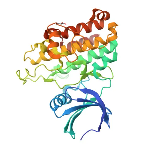
- TGF-beta receptor type-2 (TGFBR2) is a transmembrane serine/threonine kinase that forms a heteromeric complex with TGFBR1 to serve as the specific receptor for TGF-beta cytokines (TGFB1, TGFB2, TGFB3).
- Upon ligand binding, TGFBR2 phosphorylates and activates TGFBR1, which then phosphorylates SMAD2, leading to formation of the SMAD2-SMAD4 complex that translocates to the nucleus to regulate gene transcription (canonical SMAD-dependent pathway). Non-canonical, SMAD-independent signaling pathways are also initiated.
- TGFBR2 plays critical roles in cell cycle arrest, mesenchymal cell proliferation and differentiation, wound healing, extracellular matrix production, immunosuppression, and carcinogenesis.
- The receptor binds TGFB1, TGFB2, and TGFB3 with picomolar affinity without additional co-receptors and can modulate SMAD2/3 activation in specific contexts.
- Subcellularly, TGFBR2 is primarily localized at the cell membrane, but is also found in membrane rafts and can be secreted.
- The protein undergoes alternative splicing, phosphorylation, and glycosylation; mutations are associated with aortic aneurysm, craniosynostosis, and hereditary nonpolyposis colorectal cancer.
Experimental Guidance and Technical Tips
- The immunogen (aa 23–130) corresponds to the N-terminal extracellular domain; thus, the antibody may detect both full-length and shed/soluble forms of TGFBR2 in WB and ELISA.
- For Western blot, consider reducing conditions and anticipate bands around 65–67 kDa for the full-length receptor. Lower molecular weight species (e.g., ~9 kDa) may represent degradation products or alternative isoforms; include appropriate protease inhibitors.
- Validate specificity using human cell lines with known TGFBR2 expression alongside negative controls (e.g., knockout cells or blocking peptides) to confirm band identity.
- In ELISA applications, this antibody can serve as a capture or detection reagent; for sandwich formats, pair with a detection antibody recognizing a non-overlapping epitope.
CamelBio: Your One-Stop Sourcing Bridge
CamelBio provides one-stop access to IVD raw materials, including validated antibody pairs, optimized monoclonal and polyclonal antibodies, and bulk ancillary reagents, supporting your TGF-beta signaling research and diagnostic development. With capabilities covering every stage from concept to clinic, we ensure reliable supply for critical targets like TGF beta Receptor II and can assist in sourcing rare target raw materials.
REQUEST A QUOTE
Our professional team will reply to you within one business day. Please feel free to contact us!