Polyclonal Antibodies
Anti-TDP1 Rabbit pAb for WB, IF/ICC, ELISA - Q9NUW8
Item Number : CM0029956
Price varies based on specs and customizations
- Application
- WB, IF/ICC, ELISA
- Cross Reactivity
- Human, Rat
- Protein Weight
- 68kDa

Shipping:
Contact us to get shipping details Enjoy On-time Dispatch Guarantee.
Why Choose Us
Easy ordering process, quality products, and dedicated support for your business success.
Core Product Specifications and Parameters
| Parameter | Value |
|---|---|
| Product Name | TDP1 Rabbit pAb |
| Remarks/Alias | TDP1 |
| Species | Human |
| Gene ID (Human) | 55775 |
| Gene ID | 55775 |
| Immunogen | Recombinant fusion protein containing a sequence corresponding to amino acids 1-140 of human TDP1 (NP_060789.2) |
| Source | Rabbit |
| Category | Polyclonal Antibodies |
| Application | WB, IF/ICC, ELISA |
| Cross Reactivity | Human, Rat |
| SWISS | Q9NUW8 |
| Protein Weight | 68kDa |
| Shipping | Ice bag |
Biological Background: TDP1 Function and Localization
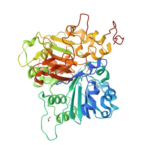
- TDP1 (tyrosyl-DNA phosphodiesterase 1) is a DNA repair enzyme that hydrolyzes 3'-phosphodiester bonds to remove covalent adducts from DNA, generating free 3' phosphate ends.
- It resolves dead-end complexes between DNA and topoisomerase I active site tyrosine residues, critical for repairing topoisomerase I-mediated DNA damage.
- Additionally, TDP1 hydrolyzes 3'-phosphoglycolates on protruding 3' ends of DNA double-strand breaks caused by radiation and free radicals, and acts on both blunt-ended double-strand breaks and single-stranded DNA.
- The enzyme possesses low 3' exonuclease activity, capable of removing a single nucleoside from the 3' end of DNA or RNA molecules with 3' hydroxyl groups, but lacks exonuclease activity on 3' phosphate-terminated substrates.
- TDP1 is encoded by the TDP1 gene (Gene ID: 55775) and belongs to the phospholipase D family, with alternative splicing producing multiple isoforms.
- It localizes to both the nucleus and cytoplasm, in line with its roles in nuclear DNA repair and potential cytoplasmic functions.
- Post-translational modifications include phosphorylation, and TDP1 is implicated in neurodegenerative disease when mutated (spinocerebellar ataxia with axonal neuropathy). Keywords: 3D-structure, Alternative splicing, Cytoplasm, Disease variant, DNA damage, DNA repair, Exonuclease, Hydrolase, Neurodegeneration, Nuclease, Nucleus, Phosphoprotein.
- Ubiquitously expressed, with similar levels throughout the central nervous system, and increased expression in testis and thymus.
Experimental Guidance and Technical Tips
- The immunogen corresponds to the N-terminal region (aa 1-140), which may be conserved across species; confirm reactivity in your species of interest.
- For Western blot, the predicted molecular weight is ~68 kDa; validate band detection in relevant cell or tissue lysates.
- In immunocytochemistry, both nuclear and cytoplasmic staining may be observed due to TDP1's dual localization; include appropriate positive and negative controls.
- ELISA can be used to assess relative TDP1 levels; consider using recombinant protein as a standard for quantification.
- As a rabbit polyclonal antibody, batch-to-batch variability is inherent; we recommend lot testing for critical applications.
CamelBio: Your One-Stop Sourcing Bridge
CamelBio is your integrated partner for IVD raw-material sourcing, providing reliable access to critical reagents from discovery to clinic. For researchers focused on DNA repair pathways or neurodegenerative disease, we can supply validated antibody pairs, monoclonal and polyclonal antibodies, and ancillary reagents to streamline your assay development. Contact us to discuss bulk orders or custom sourcing solutions for TDP1 and related targets.
REQUEST A QUOTE
Our professional team will reply to you within one business day. Please feel free to contact us!