Polyclonal Antibodies
Anti-TBK1/NAK Rabbit Polyclonal Antibody for WB, IHC-P, ELISA - Q9UHD2
Item Number : CM0030408
Price varies based on specs and customizations
- Application
- WB, IHC-P, ELISA
- Cross Reactivity
- Human, Mouse, Rat
- Protein Weight
- 84kDa

Shipping:
Contact us to get shipping details Enjoy On-time Dispatch Guarantee.
Why Choose Us
Easy ordering process, quality products, and dedicated support for your business success.
Core Product Specifications and Parameters
| Parameter | Specification |
|---|---|
| Product Name | TBK1/NAK Rabbit pAb |
| Remarks/Alias | NAK; T2K; IIAE8; FTDALS4; TBK1/NAK |
| Species | Human |
| GeneID | 29110 |
| Immunogen | Synthetic peptide corresponding to a sequence within amino acids 500-600 of human TBK1/NAK (NP_037386.1) |
| Source | Rabbit |
| Category | Polyclonal Antibodies |
| Application | WB, IHC-P, ELISA |
| Cross Reactivity | Human, Mouse, Rat |
| SWISS | Q9UHD2 |
| Protein Weight | 84kDa |
| Shipping | Ice bag |
Biological Background: TBK1 Function and Localization
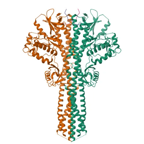
- Core innate immune kinase: Upon toll-like receptor activation by microbial components, TBK1 associates with TRAF3 and TANK, leading to phosphorylation of interferon regulatory factors IRF3 and IRF7, which triggers their dimerization, nuclear translocation, and production of type I interferons (IFNA/IFNB).
Related references: PMID:12692549, PMID:12702806, PMID:14703513, PMID:15367631, PMID:25636800 - Licensing kinase for IRF3: Phosphorylates innate adapter proteins (MAVS, STING1, TICAM1) on their pLxIS motifs, creating a docking site that recruits IRF3, thereby licensing it for subsequent phosphorylation by TBK1.
Related references: PMID:25636800, PMID:30842653, PMID:37926288 - Autophagy regulation: Restricts bacterial proliferation by phosphorylating the autophagy receptor OPTN (Ser177) to enhance LC3 binding, and promotes autophagosome maturation via phosphorylation of SMCR8 and ATG8 proteins (MAP1LC3C, GABARAPL2).
Related references: PMID:21617041, PMID:27103069, PMID:31709703 - NF-κB pathway modulation: Under certain conditions, TBK1 can activate NF-κB signaling by phosphorylating NFKBIA (IκBα), IKBKB, or RELA, leading to nuclear translocation of NF-κB.
Related references: PMID:10783893, PMID:15489227 - Expression and localization: Ubiquitously expressed with highest levels in testis; also found in retinal ganglion cells, nerve fiber layer, and microvasculature. Subcellularly localized to the cytoplasm.
- Disease relevance: TBK1 mutations are linked to amyotrophic lateral sclerosis (FTDALS4) and glaucoma, highlighting its neuropathological and ophthalmological significance.
Experimental Guidance and Technical Tips
- For Western blotting, TBK1 migrates at ~84 kDa; include a loading control of similar molecular weight and validate detection in cell lines of human, mouse, or rat origin (e.g., immune cells or retinal tissues).
- Consider stimulating cells with viral mimetics (poly(I:C)) or TLR agonists to enhance endogenous TBK1 signal.
- For IHC-P, antigen retrieval optimization is recommended; test sections of testis or retina as positive controls based on high expression levels.
- The immunogen (a.a. 500–600) is in the C-terminal region; ensure sample preparation does not mask or cleave this epitope.
- ELISA applications may require a pair with another TBK1 antibody; consider testing in sandwich format with recombinant protein.
CamelBio: Your One-Stop Sourcing Bridge
CamelBio provides diagnostic manufacturers, labs, and research institutes with one-stop access to IVD raw materials, technical services, and consulting—covering every stage from concept to clinic. For immunology and infectious disease IVD development, we offer validated antibody pairs, optimized monoclonal/polyclonal antibodies, bulk ancillary reagents, and rare target raw-material sourcing expertise. Whether you need this anti-TBK1/NAK antibody or custom solutions for kinase signaling pathways, CamelBio bridges your procurement and development needs.
Product Datasheet
Anti-TBK1/NAK Rabbit Polyclonal Antibody for WB, IHC-P, ELISA - Q9UHD2
REQUEST A QUOTE
Our professional team will reply to you within one business day. Please feel free to contact us!