Monoclonal Antibodies
Anti-TBK1/NAK Monoclonal Antibody (KO Validated) for WB - Q9UHD2
Item Number : CM0012383
Price varies based on specs and customizations
- Application
- WB, IF/ICC, IHC-P, IP, ELISA
- Cross Reactivity
- Human, Mouse, Rat
- Protein Weight
- 84 kDa

Shipping:
Contact us to get shipping details Enjoy On-time Dispatch Guarantee.
Why Choose Us
Easy ordering process, quality products, and dedicated support for your business success.
Core Product Specifications and Parameters
| Parameter | Value |
|---|---|
| Product Name | [KO Validated] TBK1/NAK Rabbit mAb |
| Remarks/Alias | NAK; T2K; IIAE8; FTDALS4; TBK1/NAK |
| Species | Human |
| GeneID (Human) | 29110 |
| GeneID | 29110 |
| Immunogen | A synthetic peptide corresponding to a sequence within amino acids 1-100 of human TBK1/NAK (Q9UHD2) |
| Source | Rabbit |
| Category | Monoclonal Antibodies |
| Application | WB, IF/ICC, IHC-P, IP, ELISA |
| Cross Reactivity | Human, Mouse, Rat |
| SWISS | Q9UHD2 |
| Protein Weight | 84 kDa |
| Shipping | Ice bag |
Biological Background: TBK1/NAK Function and Localization
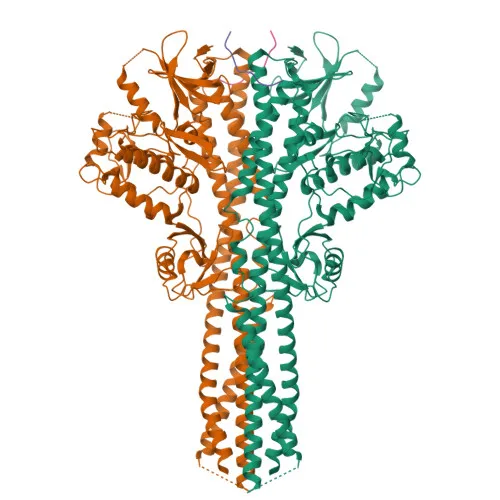
- TBK1 (also known as NAK or T2K) is a serine/threonine kinase essential for regulating inflammatory responses against pathogens. Related references: PMID:10581243, PMID:11839743, PMID:12692549
- Upon TLR activation, TBK1 associates with TRAF3 and TANK, phosphorylating IRF3 and IRF7 to induce antiviral genes. Related references: PMID:12692549, PMID:12702806, PMID:14703513, PMID:15367631, PMID:25636800
- TBK1 phosphorylates adapter proteins (MAVS, STING1, TICAM1) on pLxIS motifs to recruit and license IRF3. Related references: PMID:25636800, PMID:30842653, PMID:37926288
- It can also activate NF-κB signaling by phosphorylating NFKBIA, IKBKB, or RELA. Related references: PMID:10783893, PMID:15489227
- TBK1 promotes antibacterial autophagy via OPTN phosphorylation and regulates autophagosome maturation through SMCR8 and ATG8 proteins. Related references: PMID:21617041, PMID:27103069, PMID:31709703
- TBK1 is ubiquitously expressed, with higher levels in testis and retina; mutations are associated with amyotrophic lateral sclerosis and glaucoma. It primarily localizes to the cytoplasm.
Experimental Guidance and Technical Tips
- The immunogen is a synthetic peptide spanning the N-terminal region (aa 1–100) of human TBK1, which is conserved across human, mouse, and rat, consistent with the antibody’s multi-species reactivity.
- For Western blotting, TBK1 migrates at approximately 84 kDa; include appropriate molecular weight markers and loading controls.
- For IF/ICC and IHC-P, validate staining patterns in known TBK1-expressing tissues (e.g., testis, retina) or cell lines, and titrate the antibody to minimize background.
- For IP and ELISA applications, optimize antibody concentration and consider using antigen-specific blocking to ensure specificity.
CamelBio: Your One-Stop Sourcing Bridge
CamelBio provides diagnostic manufacturers and research institutes with a comprehensive one-stop platform for IVD raw materials. From validated antibody pairs and optimized monoclonal antibodies to bulk ancillary reagents, we help you source everything needed for your assay development. Our anti-TBK1/NAK monoclonal antibody supports research in innate immunity, inflammation, autophagy, and neurodegenerative diseases. Contact us to streamline your procurement and accelerate your project from concept to clinic.
REQUEST A QUOTE
Our professional team will reply to you within one business day. Please feel free to contact us!