Monoclonal Antibodies
Anti-Talin 2 Rabbit Monoclonal Antibody for WB, IF-P, IHC-P, ELISA - Q9Y4G6
Item Number : CM0003292
Price varies based on specs and customizations
- Application
- WB, IF-P, IHC-P, ELISA
- Cross Reactivity
- Human, Mouse, Rat
- Protein Weight
- 272kDa

Shipping:
Contact us to get shipping details Enjoy On-time Dispatch Guarantee.
Why Choose Us
Easy ordering process, quality products, and dedicated support for your business success.
Core Product Specifications and Parameters
| Parameter | Value |
|---|---|
| Product Name | Talin 2 Rabbit mAb |
| Remarks/Alias | ILWEQ, TLN2, Talin-2 |
| Species | Human |
| Gene ID (Human) | 83660 |
| Immunogen | Synthetic peptide corresponding to a sequence within amino acids 350-450 of human Talin 2 (Q9Y4G6) |
| Source | Rabbit |
| Category | Monoclonal Antibodies |
| Application | WB, IF-P, IHC-P, ELISA |
| Cross Reactivity | Human, Mouse, Rat |
| SWISS | Q9Y4G6 |
| Protein Weight | 272kDa |
| Shipping | Ice bag |
Biological Background: Talin-2 Function and Localization
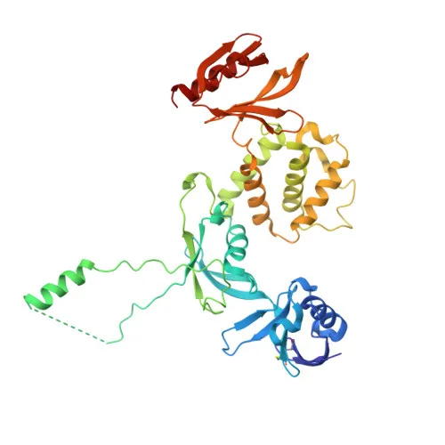
- Talin-2 (encoded by TLN2) is a large (~272 kDa) cytoskeletal scaffold protein and a core component of focal adhesion plaques.
- It functions as a critical linker between integrin adhesion receptors and the actin cytoskeleton, regulating cell adhesion, spreading, and migration.
- Talin-2 recruits phosphatidylinositol-4-phosphate 5-kinase type-1 gamma (PIP5K1C) to focal adhesions and potently activates its kinase activity, thereby modulating local PIP2 levels and downstream signaling.
- Subcellularly, Talin-2 localizes to the cytoplasm, cell membrane, and specialized adhesion structures such as focal adhesions and synapses; it also associates with the actin cytoskeleton.
- Post‑translational modifications include phosphorylation (phosphoprotein), and its three‑dimensional structure has been resolved (3D‑structure keyword).
- Talin-2 is part of the human reference proteome and has been identified in multiple proteomics studies, highlighting its ubiquitous and functional importance.
Experimental Guidance and Technical Tips
- The immunogen lies within amino acids 350–450 of human Talin-2; therefore, the antibody may detect N‑terminal truncated isoforms. Validate the target band(s) in your sample.
- For western blotting, use a low‑percentage SDS‑PAGE gel (e.g., 6–8%) and extended transfer times to ensure efficient detection of the high‑molecular‑weight (272 kDa) Talin‑2 protein.
- In immunofluorescence and immunohistochemistry, include a permeabilization step, as Talin‑2 is a cytosolic and cytoskeleton‑associated protein. Colocalization with focal adhesion markers (e.g., vinculin, paxillin) is recommended.
- The antibody cross‑reacts with human, mouse, and rat samples; however, performance should be confirmed in the specific experimental context (e.g., cell type, tissue fixation conditions).
- For ELISA applications, pair the antibody with a matched detection reagent (e.g., an anti‑Talin‑2 polyclonal antibody) after validating binding linearity and specificity.
CamelBio: Your One-Stop Sourcing Bridge
CamelBio provides diagnostic manufacturers, labs, and research institutes with a reliable, one‑stop channel for IVD raw materials, technical services, and consulting. For researchers investigating cell adhesion, focal adhesion dynamics, and cytoskeletal organization, we supply validated antibody pairs, optimized monoclonal/polyclonal antibodies (including rabbit monoclonal Talin‑2 antibodies), bulk ancillary reagents, and can assist in sourcing rare‑target raw materials. Whether you need a single reagent or an integrated supply solution from concept to clinic, contact us to streamline your procurement and development.
REQUEST A QUOTE
Our professional team will reply to you within one business day. Please feel free to contact us!