Polyclonal Antibodies
Anti-TAK1 Polyclonal Antibody for WB, IHC-P, IF/ICC, ELISA - O43318
Item Number : CM0017161
Price varies based on specs and customizations
- Application
- WB, IHC-P, IF/ICC, ELISA
- Cross Reactivity
- Human, Mouse, Rat
- Protein Weight
- 67kDa

Shipping:
Contact us to get shipping details Enjoy On-time Dispatch Guarantee.
Why Choose Us
Easy ordering process, quality products, and dedicated support for your business success.
Core Product Specifications and Parameters
| Parameter | Value |
|---|---|
| Product Name | TAK1 Rabbit pAb |
| Remarks/Alias | CSCF; FMD2; TAK1; MEKK7; TGF1a |
| Species | Human |
| GeneID (Human) | 6885 |
| GeneID | 6885 |
| Immunogen | Synthetic peptide corresponding to a sequence within amino acids 507-606 of human TAK1 (O43318) |
| Source | Rabbit |
| Category | Polyclonal Antibodies |
| Application | WB, IHC-P, IF/ICC, ELISA |
| Cross Reactivity | Human, Mouse, Rat |
| SWISS | O43318 |
| Protein Weight | 67kDa |
| Shipping | Ice bag |
Biological Background: TAK1 Function and Localization
-
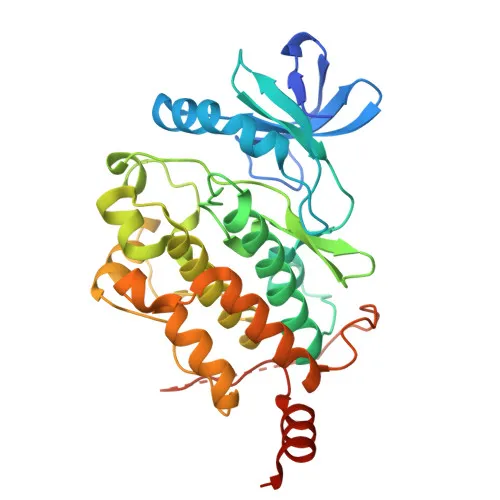
TAK1 is a serine/threonine kinase that serves as an essential component of the MAP kinase signal transduction pathway. Related references: PMID:10094049 PMID:11460167 PMID:12589052
-
It mediates signal transduction from various stimuli including interleukin-1 (IL-1), transforming growth factor-beta (TGFB), TGFB-related factors (BMP2, BMP4), toll-like receptors (TLR), CD40, and B-cell receptor (BCR). Related references: PMID:16893890 PMID:9079627
-
TAK1 activates the MKK/JNK and p38 MAPK cascades by phosphorylating MAP2Ks like MAP2K1/MEK1, MAP2K3/MKK3, MAP2K6/MKK6, and MAP2K7/MKK7, leading to AP-1 transcription factor regulation. It also activates NF-kappa-B by promoting I-kappa-B-kinase (IKK) complex activation independently of MAP2Ks. Related references: PMID:11460167 PMID:8663074
-
TAK1 promotes TRIM5 capsid-specific restriction activity and phosphorylates STING1 in response to cGAMP-activation, promoting STING1 translocation to COPII vesicles. Related references: PMID:21512573 PMID:37832545
-
TAK1 is found in the cytoplasm and at the cell membrane.
-
Multiple isoforms are expressed in a tissue-specific manner: isoform 1A is abundant in ovary, skeletal muscle, spleen, and blood mononuclear cells; 1B in brain, kidney, small intestine; 1C in prostate; 1D is less abundant.
-
TAK1 is subject to acetylation, phosphorylation, and ubiquitination. It is an ATP-binding, magnesium-dependent kinase involved in stress response, transcription regulation, and apoptosis.
Experimental Guidance and Technical Tips
- The immunogen corresponds to the C-terminal region (aa 507-606) of human TAK1. Consider verifying reactivity against your specific isoform of interest.
- For Western blotting, expect a band around 67 kDa. For immunohistochemistry and immunofluorescence, optimize antigen retrieval and permeabilization conditions.
- The antibody cross-reacts with mouse and rat, enabling studies in these common model organisms. Validate in the relevant sample system.
- ELISA applications may require pairing with a suitable secondary antibody; consider testing dilution ranges experimentally.
CamelBio: Your One-Stop Sourcing Bridge
CamelBio provides diagnostic manufacturers and research labs with a streamlined pathway to high-quality IVD raw materials. This anti-TAK1 polyclonal antibody is part of our comprehensive portfolio for signal transduction and inflammation research, supporting projects from early development to clinical application. Contact us for bulk custom antibody production, validated antibody pairs, and ancillary reagents tailored to your assay needs.
REQUEST A QUOTE
Our professional team will reply to you within one business day. Please feel free to contact us!