Polyclonal Antibodies
Anti-SUGCT Polyclonal Antibody for WB, ELISA - Q9HAC7
Item Number : CM0026100
Price varies based on specs and customizations
- Application
- WB, ELISA
- Cross Reactivity
- Human, Mouse, Rat
- Protein Weight
- 48kDa

Shipping:
Contact us to get shipping details Enjoy On-time Dispatch Guarantee.
Why Choose Us
Easy ordering process, quality products, and dedicated support for your business success.
Core Product Specifications and Parameters
| Parameter | Value |
|---|---|
| Product Name | SUGCT Rabbit pAb |
| Remarks/Alias | GA3; ORF19; DERP13; C7orf10; SUGCT |
| Species | Human |
| GeneID (human) | 79783 |
| GeneID | 79783 |
| Immunogen | Recombinant protein|Recombinant fusion protein containing a sequence corresponding to amino acids 1-250 of human SUGCT (NP_001180240.1). |
| Source | Rabbit |
| Category | Polyclonal Antibodies |
| Application | WB, ELISA |
| Cross Reactivity | Human, Mouse, Rat |
| SWISS | Q9HAC7 |
| Protein Weight | 48kDa |
| Shipping | Ice bag |
Biological Background: SUGCT Function and Localization
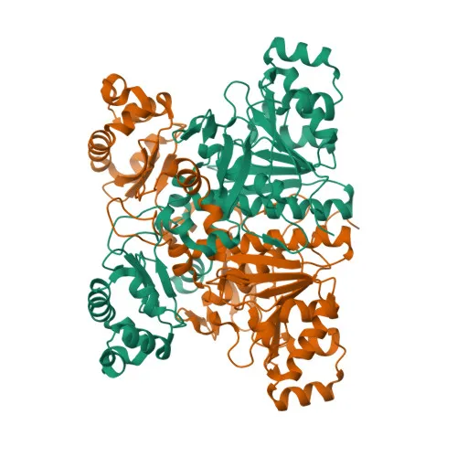
- SUGCT (Succinyl-CoA:glutarate CoA-transferase) is a mitochondrial enzyme encoded by the SUGCT gene, also known as Dermal papilla-derived protein 13 (DERP13), with a molecular weight of ~48 kDa.
- It catalyzes the reversible transfer of CoA from dicarboxyl-CoAs (preferring succinyl-CoA and glutaryl-CoA) to dicarboxylates including glutarate, succinate, adipate, itaconate, and 3-hydroxy-3-methylglutarate, playing a key role in metabolite recycling (PubMed:23893049, PubMed:34492704, PubMed:38915184). Related references: PMID:23893049, PMID:34492704, PMID:38915184
- SUGCT acts in the lysine and tryptophan degradation pathway, catalyzing succinyl-CoA-dependent reesterification of free glutarate to glutaryl-CoA to prevent renal loss (PubMed:18926513, PubMed:23893049, PubMed:38915184). Related references: PMID:18926513, PMID:23893049, PMID:38915184
- During inflammation, SUGCT may convert macrophage-derived itaconate to itaconyl-CoA in erythroid precursors, thereby negatively regulating the TCA cycle, heme synthesis, and stress erythropoiesis (PubMed:34492704). Related references: PMID:34492704
- Immunofluorescence and electron microscopy studies localize SUGCT predominantly to mitochondria.
- Tissue expression analysis shows high expression in kidney, with moderate levels in liver, skeletal muscle, and pancreas, and low/absent expression elsewhere.
- UniProt keywords highlight the protein as a transferase with a transit peptide, acetylation sites, and involvement in glutaric aciduria pathology; alternative splicing and disease variants are documented.
Experimental Guidance and Technical Tips
- The immunogen spans the N-terminal 250 amino acids of human SUGCT, potentially enabling detection of full-length and processed forms; consider the transit peptide cleavage site when assessing band sizes in Western blotting.
- For Western blotting, the predicted molecular weight is ~48 kDa (precursor) or ~45 kDa (mature); validate in lysates from kidney or liver tissues where expression is high.
- For ELISA, optimize coating concentrations and validate in the relevant sample matrix; cross-reactivity with mouse and rat orthologs should be confirmed by sequence comparison.
CamelBio: Your One-Stop Sourcing Bridge
CamelBio serves as a comprehensive IVD raw-material sourcing partner, connecting diagnostic manufacturers and research teams with high-quality antibodies like this anti-SUGCT polyclonal antibody for metabolism and disease research. Our portfolio includes validated antibody pairs, optimized monoclonal/polyclonal antibodies, bulk ancillary reagents, and custom raw-material sourcing for rare targets, streamlining your development from concept to clinic.
REQUEST A QUOTE
Our professional team will reply to you within one business day. Please feel free to contact us!