Polyclonal Antibodies
Anti-SPSB1 Polyclonal Antibody for WB and ELISA - Q96BD6
Item Number : CM0015199
Price varies based on specs and customizations
- Application
- WB, ELISA
- Cross Reactivity
- Human, Mouse, Rat
- Protein Weight
- 31kDa

Shipping:
Contact us to get shipping details Enjoy On-time Dispatch Guarantee.
Why Choose Us
Easy ordering process, quality products, and dedicated support for your business success.
Core Product Specifications and Parameters
| Parameter | Value |
|---|---|
| Product Name | SPSB1 Rabbit pAb |
| Remarks/Alias | SSB1; SSB-1; SPSB1 |
| Species | Human |
| GeneID (human) | 80176 |
| GeneID | 80176 |
| Immunogen | Recombinant fusion protein containing a sequence corresponding to amino acids 1-60 of human SPSB1 (NP_079382.2). |
| Source | Rabbit |
| Category | Polyclonal Antibodies |
| Application | WB, ELISA |
| Cross Reactivity | Human, Mouse, Rat |
| SWISS | Q96BD6 |
| Protein Weight | 31kDa |
| Shipping | Ice bag |
Biological Background: SPSB1 Function and Localization
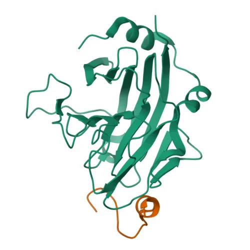
- SPSB1 (SPRY domain-containing SOCS box protein 1), also known as SSB1 and SSB-1, is a member of the SPRY domain-containing SOCS box protein family.
- It functions as the substrate recognition component of an SCF-like ECS (Elongin BC-CUL2/5-SOCS-box protein) E3 ubiquitin-protein ligase complex, mediating the ubiquitination and subsequent proteasomal degradation of target proteins. Related references: PMID:15601820 PMID:21199876
- SPSB1 negatively regulates nitric oxide (NO) production and limits cellular toxicity in activated macrophages by mediating the ubiquitination and proteasomal degradation of NOS2. Related references: PMID:21199876
- Acts as a bridge which links NOS2 with the ECS E3 ubiquitin ligase complex components ELOC and CUL5.
- Localizes predominantly to the cytoplasm and cytosol.
- Post-translational modifications include phosphorylation (Phosphoprotein).
- Keywords: 3D-structure, Cytoplasm, Phosphoprotein, Proteomics identification, Reference proteome, Ubl conjugation pathway.
Experimental Guidance and Technical Tips
- The immunogen corresponds to the N-terminal region (aa 1-60) of human SPSB1; verify that this epitope is conserved in mouse and rat for expected cross-reactivity.
- For Western blot (WB), consider starting with a dilution range of 1:500–1:2000 and optimize based on signal-to-noise ratio in your sample system.
- For ELISA, test the antibody as a capture or detection reagent in a suitable sandwich or direct format; titrate to determine optimal working concentration.
- Validate detection in activated macrophage cell lines (e.g., RAW 264.7) or tissues where SPSB1-mediated regulation of NOS2 is relevant; include untreated controls to assess specificity.
- Due to the polyclonal nature, assess lot-to-lot consistency and re-validate critical assays when using a new batch.
CamelBio: Your One-Stop Sourcing Bridge
CamelBio provides diagnostic manufacturers, laboratories, and research institutes with a one-stop sourcing solution for IVD raw materials, technical services, and consulting—supporting every stage from concept to clinic. This anti-SPSB1 polyclonal antibody targets a key player in the ubiquitin-proteasome system and nitric oxide signaling, making it a valuable tool for research in inflammation, macrophage biology, and related diagnostic assay development. CamelBio can support your projects with validated antibody pairs, optimized monoclonal and polyclonal antibodies, bulk ancillary reagents, and specialized raw-material sourcing for rare or novel targets.
REQUEST A QUOTE
Our professional team will reply to you within one business day. Please feel free to contact us!