Polyclonal Antibodies
Anti-SPK (TOPK) Rabbit Polyclonal Antibody - Q96KB5
Item Number : CM0030411
Price varies based on specs and customizations
- Application
- WB, IHC-P, ELISA
- Cross Reactivity
- Human, Rat
- Protein Weight
- 36kDa

Shipping:
Contact us to get shipping details Enjoy On-time Dispatch Guarantee.
Why Choose Us
Easy ordering process, quality products, and dedicated support for your business success.
Core Product Specifications and Parameters
| Parameter | Value |
|---|---|
| Product Name | SPK Rabbit pAb |
| Remarks/Alias | SPK; CT84; TOPK; HEL164; Nori-3 |
| Species | Human |
| GeneID (Human) | 55872 |
| GeneID | 55872 |
| Immunogen | Recombinant fusion protein containing a sequence corresponding to amino acids 1-322 of human SPK (NP_060962.2). |
| Source | Rabbit |
| Category | Polyclonal Antibodies |
| Application | WB, IHC-P, ELISA |
| Cross Reactivity | Human, Rat |
| SWISS | Q96KB5 |
| Protein Weight | 36kDa |
| Shipping | Ice bag |
Biological Background: SPK (TOPK) Function and Localization
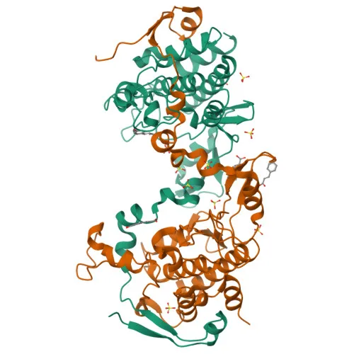
- SPK, also known as TOPK (Lymphokine-activated killer T-cell-originated protein kinase), CT84, Nori-3, PDZ-binding kinase, or MAPKK-like protein kinase, is a serine/threonine-protein kinase encoded by the PBK gene in humans.
- It functions as a mitotic kinase, phosphorylating MAP kinase p38 and appearing to be active primarily during mitosis.
- SPK may also play a role in lymphoid cell activation.
- Upon phosphorylation, SPK forms a complex with TP53, resulting in TP53 destabilization and attenuation of the G2/M checkpoint following doxorubicin-induced DNA damage.
- SPK is classified as a cancer/testis antigen (CT84) and is restrictedly expressed in testis (outer cell layer of seminiferous tubules) and placenta.
- The protein contains an ATP-binding site and undergoes post‑translational modifications including phosphorylation, acetylation, and ubiquitination‑like conjugation.
Experimental Guidance and Technical Tips
- The immunogen spans the N‑terminal region (amino acids 1‑322) of human SPK, enabling recognition of full‑length SPK and potential isoforms; verify specificity in your experimental system.
- For Western blot, consider testing antibody dilutions in the range of 1:500–1:2000 and use positive controls such as testis or placenta lysates that naturally express SPK.
- In IHC-P, antigen retrieval conditions may be necessary; optimize according to your tissue type and fixation protocol.
- When working with rat samples, confirm cross‑reactivity in a pilot experiment, as reactivity is supplier‑indicated but may require validation.
- ELISA applications may benefit from optimizing coating buffer, blocking conditions, and antibody concentration to achieve the desired sensitivity.
CamelBio: Your One‑Stop Sourcing Bridge
CamelBio empowers diagnostic manufacturers, laboratories, and research institutes with streamlined access to IVD raw materials, technical services, and consultative support across the development pipeline. This anti‑SPK (TOPK) antibody supports discovery in oncology, cell cycle regulation, and signal transduction. Through our integrated supply network, we offer validated antibody pairs, optimized monoclonal/polyclonal antibodies, bulk ancillary reagents, and rare‑target raw‑material sourcing to accelerate your project from concept to clinic.
REQUEST A QUOTE
Our professional team will reply to you within one business day. Please feel free to contact us!