Polyclonal Antibodies
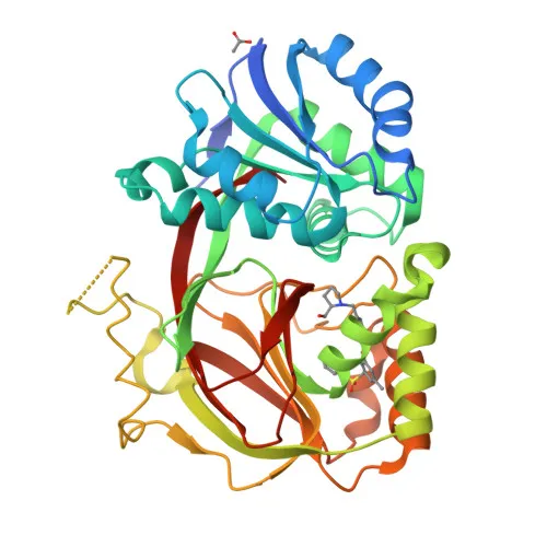
Anti-SPHK1 Rabbit Polyclonal Antibody for WB and ELISA - Q9NYA1
Item Number : CM0018660
Price varies based on specs and customizations
- Application
- WB, ELISA
- Cross Reactivity
- Human, Mouse
- Protein Weight
- 43 kDa

Shipping:
Contact us to get shipping details Enjoy On-time Dispatch Guarantee.
Why Choose Us
Easy ordering process, quality products, and dedicated support for your business success.
Core Product Specifications and Parameters
| Parameter | Value |
|---|---|
| Product Name | SPHK1 Rabbit pAb |
| Remarks/Alias | SPHK; SPHK1 |
| Species | Human |
| GeneID (Human) | 8877 |
| GeneID | 8877 |
| Immunogen | Recombinant fusion protein containing a sequence corresponding to amino acids 50-384 of human SPHK1 (NP_001136074.1) |
| Source | Rabbit |
| Category | Polyclonal Antibodies |
| Application | WB, ELISA |
| Cross Reactivity | Human, Mouse |
| SWISS | Q9NYA1 |
| Protein Weight | 43 kDa |
| Shipping | Ice bag |
Biological Background: Sphingosine Kinase 1 Function and Localization
- SPHK1 catalyzes the phosphorylation of sphingosine to form sphingosine-1-phosphate (S1P), a lipid mediator with intra- and extracellular functions. It also acts on D-erythro-sphingosine and, to a lesser extent, sphinganine, but not on other lipids such as diacylglycerol, ceramide, or phosphatidylinositol. Related references: PMID:11923095 PMID:20577214 PMID:23602659
- In contrast to proapoptotic SPHK2, SPHK1 has a negative effect on intracellular ceramide levels, enhances cell growth, and inhibits apoptosis. Related references: PMID:16118219
- Via its product S1P, SPHK1 stimulates TRAF2 E3 ubiquitin ligase activity and promotes activation of NF-kappa-B in response to TNF signaling, leading to IL17 secretion. Related references: PMID:20577214
- In response to TNF, SPHK1 negatively regulates RANTES induction through the p38 MAPK signaling pathway. Related references: PMID:23935096
- SPHK1 is involved in endocytic membrane trafficking, recruited to dilated endosomes, and plays a role in later stages of endosomal maturation and membrane fusion independently of its kinase activity. Related references: PMID:24929359 PMID:28049734
- In Purkinje cells, SPHK1 is involved in the regulation of autophagosome-lysosome fusion upon VEGFA. Related references: PMID:25417698
- SPHK1 also exhibits serine acetyltransferase activity on PTGS2/COX2 in an acetyl-CoA-dependent manner, modulating neuroinflammation by promoting neuronal secretion of specialized pro-resolving mediators.
- Subcellular localization includes cytoplasm, nucleus, cell membrane, endosome membrane, clathrin-coated pits, and synapse.
- Widely expressed, with highest levels in adult liver, kidney, heart, and skeletal muscle; also expressed in brain cortex. Related references: PMID:29662056
Experimental Guidance and Technical Tips
- The immunogen is a recombinant protein spanning amino acids 50-384 of human SPHK1, which includes the catalytic domain. For Western blot, consider a predicted molecular weight of approximately 43 kDa. Validate antibody specificity using appropriate human or mouse tissue lysates, as cross-reactivity is reported for both species.
- For ELISA, optimize antibody coating concentration and assay conditions. Consider using recombinant SPHK1 protein as a positive control.
- Since SPHK1 is involved in multiple signaling pathways, verify detection under relevant stimulation conditions (e.g., TNF treatment) to assess functional context.
CamelBio: Your One-Stop Sourcing Bridge
CamelBio is a one-stop IVD raw-material sourcing partner dedicated to connecting diagnostic manufacturers, laboratories, and research institutes with high-quality antibodies and ancillary reagents. For studies targeting SPHK1 in cell signaling, inflammation, and lipid metabolism, we provide access to validated polyclonal and monoclonal antibodies, optimized antibody pairs, and bulk reagents to support your assay development from concept to clinic.
REQUEST A QUOTE
Our professional team will reply to you within one business day. Please feel free to contact us!