Monoclonal Antibodies
Anti-SMPD1 (KO Validated) Rabbit Monoclonal Antibody for WB, IF-F, IHC-P, ELISA - P17405
Item Number : CM0007145
Price varies based on specs and customizations
- Application
- WB, IF-F, IHC-P, ELISA
- Cross Reactivity
- Human, Rat
- Protein Weight
- 70 kDa

Shipping:
Contact us to get shipping details Enjoy On-time Dispatch Guarantee.
Why Choose Us
Easy ordering process, quality products, and dedicated support for your business success.
Core Product Specifications and Parameters
| Parameter | Value |
|---|---|
| Product Name | Anti-SMPD1 (KO Validated) Rabbit Monoclonal Antibody |
| Remarks/Alias | ASM; NPD; ASMASE; D1 |
| Species | Human |
| Gene ID (Human) | 6609 |
| Gene ID | 6609 |
| Immunogen | Recombinant fusion protein containing a sequence corresponding to amino acids 70-340 of human SMPD1 (NP_000534.3) |
| Source | Rabbit |
| Category | Monoclonal Antibodies |
| Application | WB, IF-F, IHC-P, ELISA |
| Cross Reactivity | Human, Rat |
| SWISS | P17405 |
| Protein Weight | 70 kDa |
| Shipping | Ice bag |
Biological Background: SMPD1 Function and Localization
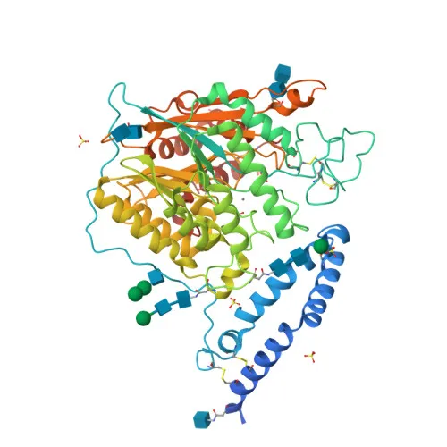
- SMPD1 encodes sphingomyelin phosphodiesterase, also known as acid sphingomyelinase (aSMase). Alternative splicing produces isoforms; the protein exists as two enzymatic forms from alternative trafficking: one endolysosomal, the other secreted. Related references: PMID:20807762 PMID:21098024 PMID:9660788
- In the lysosomes, aSMase converts sphingomyelin to ceramide, playing a critical role in cholesterol export from intraendolysosomal membranes and exhibiting phospholipase C activities. Related references: PMID:20807762 PMID:21098024 PMID:25339683
- Modulates stress-induced apoptosis through the production of ceramide. Related references: PMID:8706124
- Secreted aSMase reorganizes plasma membrane by converting sphingomyelin to ceramide, and its secretion is upregulated by inflammatory mediators and pathogens, facilitating host defense and pathogen internalization. Related references: PMID:12563314 PMID:17303575 PMID:20807762 PMID:9393854
- During bacterial infection, the lysosomal form is released extracellularly and cleaved by CASP7, promoting plasma membrane repair and counteracting GSDMD perforin pores. Related references: PMID:21157428
- Subcellular localization includes lysosomes, lipid droplets, and secretion into the extracellular space.
- Post-translational modifications include glycosylation, phosphorylation, and disulfide bond formation; the enzyme is zinc-dependent (metal-binding).
Experimental Guidance and Technical Tips
- The antibody is raised against a recombinant fragment encompassing amino acids 70–340 of human SMPD1, which may recognize epitopes within the catalytic domain. Consider validating specificity using knockout (KO) cell lysates where available.
- For Western blotting (WB), use reducing conditions and expect a band at approximately 70 kDa. Lysosomal enrichment or treatment with stressors may enhance detection.
- In immunofluorescence (IF-F) and immunohistochemistry (IHC-P), permeabilization and antigen retrieval are recommended. Co-staining with lysosomal markers can confirm subcellular localization.
- For ELISA, the antibody may be used as part of a matched pair; titrate and optimize conditions for your specific assay format.
- Cross-reactivity is reported for human and rat; for other species, validation is advised.
CamelBio: Your One-Stop Sourcing Bridge
CamelBio is a one-stop provider of IVD raw materials, supporting diagnostic developers and researchers with reliable antibodies, antigens, and custom sourcing. This anti-SMPD1 rabbit monoclonal antibody serves as a key tool for studying sphingolipid metabolism, lysosomal storage disorders, and pathogen-host interactions. Beyond this product, CamelBio offers validated antibody pairs, bulk reagents, and sourcing for rare targets to streamline your assay development from concept to clinic.
Product Datasheet
Anti-SMPD1 (KO Validated) Rabbit Monoclonal Antibody for WB, IF-F, IHC-P, ELISA - P17405
REQUEST A QUOTE
Our professional team will reply to you within one business day. Please feel free to contact us!