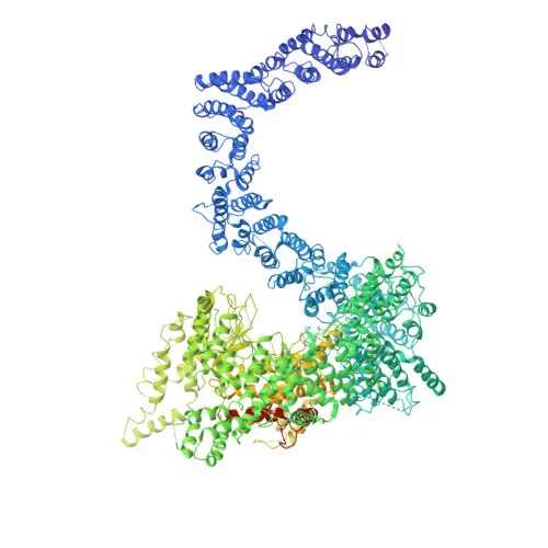
Monoclonal Antibodies
Anti-SMG1 Rabbit Monoclonal Antibody for WB and ELISA - Q96Q15
Item Number : CM0002672
Price varies based on specs and customizations
- Application
- WB, ELISA
- Cross Reactivity
- Mouse
- Protein Weight
- 411 kDa

Shipping:
Contact us to get shipping details Enjoy On-time Dispatch Guarantee.
Why Choose Us
Easy ordering process, quality products, and dedicated support for your business success.
Core Product Specifications and Parameters
| Parameter | Value |
|---|---|
| Product Name | SMG1 Rabbit mAb |
| Remarks/Alias | ATX; LIP; 61E3.4; SMG1 |
| Species | Human |
| GeneID (Human) | 23049 |
| GeneID | 23049 |
| Immunogen | Synthetic peptide|A synthetic peptide corresponding to a sequence within amino acids 3562-3661 of human SMG1 (Q96Q15). |
| Source | Rabbit |
| Category | Monoclonal Antibodies |
| Application | WB, ELISA |
| Cross Reactivity | Mouse |
| SWISS | Q96Q15 |
| Protein Weight | 411 kDa |
| Shipping | Ice bag |
Biological Background: SMG1 Function and Localization
- SMG1 is a serine/threonine-protein kinase that plays a central role in nonsense-mediated mRNA decay (NMD) by phosphorylating UPF1/RENT1. It is part of the SMG1C complex together with SMG8 and SMG9.
- It is recruited to stalled ribosomes to form the SURF complex (SMG1-UPF1-eRF1-eRF3), which associates with the exon junction complex (EJC) via UPF2 to trigger NMD.
- Besides NMD, SMG1 also functions in the genotoxic stress response, phosphorylating p53/TP53 and contributing to optimal p53 activation after DNA damage. Its depletion leads to spontaneous DNA damage and increased sensitivity to ionizing radiation.
- SMG1 localizes to both the nucleus and cytoplasm.
- It is widely expressed, with highest levels in heart and skeletal muscle, and also present in placenta, brain, lung, and spleen, but absent in liver.
- The protein contains an ATP-binding domain and requires manganese as a metal cofactor. It undergoes acetylation and phosphorylation.
- Alternative splicing generates multiple isoforms of SMG1.
Experimental Guidance and Technical Tips
- The immunogen corresponds to a sequence within the C-terminal region (aa 3562–3661) of human SMG1, distinct from the kinase domain, potentially allowing detection of both full-length and truncated isoforms in Western blot.
- For Western blot, due to the large protein size (~411 kDa), use a low-percentage SDS-PAGE gel (e.g., 6% or gradient) and extended transfer times to ensure efficient transfer of the high-molecular-weight protein.
- Validate antibody performance in mouse samples if cross-reactivity is required, as the antibody is reported to cross-react with mouse SMG1.
- For ELISA, consider using the same peptide as a positive control antigen to establish assay conditions.
- In NMD or DNA damage studies, use appropriate controls (e.g., UPF1 phosphorylation status) to confirm SMG1 activity in your experimental system.
CamelBio: Your One-Stop Sourcing Bridge
CamelBio provides diagnostic manufacturers and research labs with streamlined access to high-quality IVD raw materials, including validated antibody pairs and optimized monoclonal antibodies like this anti-SMG1 clone for NMD and DNA damage research. Our one-stop platform also supports bulk ancillary reagents and rare target sourcing, helping you accelerate assay development from concept to clinic.
REQUEST A QUOTE
Our professional team will reply to you within one business day. Please feel free to contact us!