Polyclonal Antibodies
Anti-SIGMAR1 Rabbit Polyclonal Antibody for WB and ELISA - Q99720
Item Number : CM0017391
Price varies based on specs and customizations
- Application
- WB, ELISA
- Cross Reactivity
- Human, Rat
- Protein Weight
- 25kDa

Shipping:
Contact us to get shipping details Enjoy On-time Dispatch Guarantee.
Why Choose Us
Easy ordering process, quality products, and dedicated support for your business success.
Core Product Specifications and Parameters
| Parameter | Value |
|---|---|
| Product Name | SIGMAR1 Rabbit pAb |
| Remarks/Alias | SRBP; ALS16; DSMA2; OPRS1; SR-BP; SIG-1R; SR-BP1; sigma1R; hSigmaR1; SIGMAR1 |
| Species | Human |
| Gene ID (Human) | 10280 |
| Immunogen | Recombinant fusion protein containing a sequence corresponding to amino acids 31-80 of human SIGMAR1 (NP_005857.1) |
| Source | Rabbit |
| Category | Polyclonal Antibodies |
| Application | WB, ELISA |
| Cross Reactivity | Human, Rat |
| SWISS | Q99720 |
| Protein Weight | 25kDa |
| Shipping | Ice bag |
Biological Background: SIGMAR1 Function and Localization
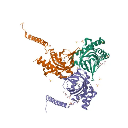
- SIGMAR1, also known as sigma non-opioid intracellular receptor 1, SRBP, or SIG1R, is a 25 kDa transmembrane protein encoded by the SIGMAR1 gene (alternative name OPRS1).
- It mediates lipid transport from the endoplasmic reticulum and regulates the biogenesis of lipid microdomains at the plasma membrane, impacting a wide array of cellular functions. Related references: PMID:16472803, PMID:9341151
- SIGMAR1 modulates multiple signaling pathways, including BDNF and EGF signaling, and regulates ion channels such as potassium channels, potentially affecting neurotransmitter release. Related references: PMID:16472803, PMID:9341151
- Together with ANK2, it controls ITP3R-dependent calcium efflux at the endoplasmic reticulum, playing a role in intracellular calcium signaling.
- The protein is necessary for proper mitochondrial axonal transport in motor neurons, particularly retrograde movement of mitochondria, and protects against oxidative stress-induced cell death via interaction with RNF112 (By similarity).
- Subcellular localization includes the endoplasmic reticulum membrane, nucleus envelope, cell membrane, cytoplasmic vesicles, lipid droplets, cell junctions, and growth cones.
- It is widely expressed, with higher levels in liver, colon, prostate, placenta, small intestine, heart, and pancreas, as well as in retinal pigment epithelial cells and alpha-motor neurons (PubMed:23314020). Related references: PMID:23314020
- Clinically, mutations in SIGMAR1 are associated with amyotrophic lateral sclerosis (ALS16) and distal spinal muscular atrophy type 2 (DSMA2), and the protein is implicated in learning, memory, and mood disorders.
Experimental Guidance and Technical Tips
- The immunogen is a recombinant protein spanning amino acids 31-80, a region that may be conserved across species; consider verifying sequence homology for your model organism.
- For Western blot, a band at approximately 25 kDa is expected in human and rat samples; optimize blocking and antibody dilution as pre-immune serum may vary between lots.
- For ELISA, the polyclonal nature allows capture-detection pairing, but cross-adsorption against relevant species may be necessary to reduce background.
- The antibody is not validated for immunohistochemistry or immunoprecipitation; if required, test under native and denaturing conditions after confirming target accessibility in your tissue of interest.
CamelBio: Your One-Stop Sourcing Bridge
CamelBio provides diagnostic manufacturers and research institutions with streamlined access to IVD raw materials, technical services, and consultancy. For SIGMAR1-focused neuroscience and cell signaling studies, we offer validated antibody pairs, optimized monoclonal and polyclonal antibodies, and bulk ancillary reagents. Reach out to us for rare target raw-material sourcing to support your assay development from concept to clinic.
REQUEST A QUOTE
Our professional team will reply to you within one business day. Please feel free to contact us!