Polyclonal Antibodies
Anti-SIGLEC5 Rabbit Polyclonal Antibody for WB and ELISA - O15389
Item Number : CM0030128
Price varies based on specs and customizations
- Application
- WB, ELISA
- Cross Reactivity
- Mouse
- Protein Weight
- 61kDa

Shipping:
Contact us to get shipping details Enjoy On-time Dispatch Guarantee.
Why Choose Us
Easy ordering process, quality products, and dedicated support for your business success.
Core Product Specifications and Parameters
| Parameter | Value |
|---|---|
| Product Name | SIGLEC5 Rabbit pAb |
| Remarks/Alias | CD170, OBBP2, CD33L2, OB-BP2, SIGLEC-5, SIGLEC5 |
| Species | Human |
| GeneID (Human) | 8778 |
| GeneID | 8778 |
| Immunogen | Recombinant fusion protein containing a sequence corresponding to amino acids 17-434 of human SIGLEC5 (NP_003821.1) |
| Source | Rabbit |
| Category | Polyclonal Antibodies |
| Application | WB, ELISA |
| Cross Reactivity | Mouse |
| SWISS | O15389 |
| Protein Weight | 61kDa |
| Shipping | Ice bag |
Biological Background: SIGLEC5 Function and Localization
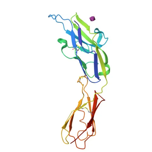
- SIGLEC5 (Sialic acid-binding Ig-like lectin 5), also known as CD170, CD33L2, and OB-BP2, is a member of the immunoglobulin superfamily and functions as a sialic acid-dependent adhesion molecule (O15389).
- It binds equally to both α-2,3-linked and α-2,6-linked sialic acid residues, though its sialic acid recognition site may be masked by cis interactions on the same cell surface.
- The protein contains immunoglobulin-like domains, a transmembrane region, and a short cytoplasmic tail, characteristic of the SIGLEC family.
- Subcellularly, SIGLEC5 is localized to the cell membrane, consistent with its role in cell-cell adhesion.
- Tissue expression is predominantly in monocytic/myeloid lineage cells, including high levels in peripheral blood leukocytes, spleen, bone marrow, and lower levels in lymph node, lung, appendix, placenta, pancreas, and thymus.
- It is expressed on monocytes and neutrophils but absent from leukemic cell lines representing early stages of myelomonocytic differentiation, indicating a role in mature myeloid cells.
- The protein contains a signal peptide, disulfide bonds, and is glycosylated, with an Ig-like V-type domain likely involved in sialic acid binding.
- Its molecular weight is approximately 61 kDa, and it is classified as a lectin with cell adhesion function.
Experimental Guidance and Technical Tips
- For Western blot, consider using protein lysates from human peripheral blood leukocytes or spleen tissue, where SIGLEC5 is highly expressed, as positive controls.
- Given the immunogen spans the extracellular domain (aa 17-434), the antibody may recognize both native and denatured forms, but optimization of blocking and incubation conditions is recommended.
- For ELISA, the antibody can be tested against recombinant SIGLEC5 protein or cell lysates from myeloid cell lines; cross-reactivity with mouse SIGLEC5 allows potential use in murine models.
- Since SIGLEC5 is glycosylated and has disulfide bonds, gentle reduction with DTT or β-mercaptoethanol may be required to maintain detection in WB; consider running reduced and non-reduced samples.
- Always validate the antibody’s specificity in your experimental system by including a negative control (e.g., knockout cell lines or siRNA knockdown samples).
CamelBio: Your One-Stop Sourcing Bridge
CamelBio is a comprehensive IVD raw-material sourcing partner supporting diagnostic developers and researchers with high-quality antibodies, including validated anti-SIGLEC5 reagents for myeloid cell adhesion studies. Our portfolio encompasses polyclonal and monoclonal antibodies, bulk ancillary reagents, and rare target sourcing to streamline your immunoassay and biomarker discovery workflows. Contact us to explore custom procurement solutions for SIGLEC5 and related CD33 family targets.
REQUEST A QUOTE
Our professional team will reply to you within one business day. Please feel free to contact us!