Monoclonal Antibodies
Anti-SERCA2/ATP2A2 Monoclonal Antibody for WB, IF/ICC, IP, ELISA - P16615
Item Number : CM0002974
Price varies based on specs and customizations
- Application
- WB, IF/ICC, IP, ELISA
- Cross Reactivity
- Human, Mouse, Rat
- Protein Weight
- 115 kDa

Shipping:
Contact us to get shipping details Enjoy On-time Dispatch Guarantee.
Why Choose Us
Easy ordering process, quality products, and dedicated support for your business success.
Core Product Specifications and Parameters
| Parameter | Value |
|---|---|
| Product Name | SERCA2/ATP2A2 Rabbit mAb |
| Remarks/Alias | DD, DAR, ATP2B, SERCA2 |
| Species | Human |
| GeneID (Human) | 488 |
| Immunogen | A synthetic peptide corresponding to a sequence within amino acids 943-1042 of human SERCA2/ATP2A2 (P16615). |
| Source | Rabbit |
| Category | Monoclonal Antibodies |
| Application | WB, IF/ICC, IP, ELISA |
| Cross Reactivity | Human, Mouse, Rat |
| SWISS | P16615 |
| Protein Weight | 115 kDa |
| Shipping | Ice bag |
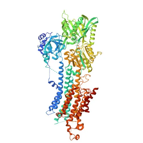
Biological Background: SERCA2/ATP2A2 Function and Localization
- SERCA2, encoded by ATP2A2, is a magnesium-dependent calcium-transporting ATPase that couples ATP hydrolysis to translocation of calcium ions from the cytosol into the sarcoplasmic/endoplasmic reticulum lumen (PubMed:12542527, PubMed:16402920). Related references: PMID:12542527 PMID:16402920
- It plays a key role in muscle contraction/relaxation by regulating cytosolic calcium levels and is essential for calcium homeostasis in non-muscle tissues.
- SERCA2 has three main isoforms generated by alternative splicing: isoform 1 widely expressed in smooth muscle and nonmuscle tissues; isoforms 2 and 3 predominantly in heart and slow-twitch skeletal muscle.
- In response to starvation, SERCA2 becomes involved in autophagy by interacting with VMP1 to control ER-isolation membrane contacts for autophagosome formation; it also modulates ER contacts with lipid droplets, mitochondria, and endosomes (PubMed:28890335). Related references: PMID:28890335
- SERCA2 is located in the endoplasmic reticulum and sarcoplasmic reticulum membranes.
- It is a critical regulator of osteoclast differentiation via TNFSF11-mediated Ca(2+) signaling, interacting with TMEM64 to drive NFATC1 activation and mitochondrial ROS production.
- Keywords include alternative splicing, calcium transport, ATP-binding, magnesium, metal-binding, transmembrane, and disulfide bond; post-translational modifications include nitration and phosphorylation.
- Tissue specificity: Isoform 1 is highest in liver, pancreas, lung; intermediate in brain, kidney, placenta; lower in heart and skeletal muscle. Isoforms 2 and 3 are cardiac/slow-twitch muscle-specific.
Experimental Guidance and Technical Tips
- The immunogen peptide (aa 943-1042) targets the C-terminal region of SERCA2. Given the high sequence similarity among isoforms, consider possible detection of multiple isoforms depending on sample type.
- For WB, an expected band around 115 kDa should be observed; validate specificity using knockout controls or siRNA knockdown experiments.
- For IF/ICC, optimize fixation and permeabilization protocols for the target cell type; note that SERCA2 localizes to ER/SR membranes, so staining may appear reticular.
- In IP and ELISA, confirm antibody performance with recombinant protein or endogenous samples; titration may be necessary to avoid high background.
CamelBio: Your One-Stop Sourcing Bridge
CamelBio serves as a comprehensive sourcing partner for diagnostic manufacturers and research labs seeking high-quality IVD raw materials. This SERCA2/ATP2A2 antibody supports investigations into calcium signaling, muscle physiology, and ER stress pathways. Beyond individual antibodies, CamelBio supplies validated antibody pairs, bulk monoclonal/polyclonal antibodies, ancillary reagents, and can assist with sourcing rare targets—helping you streamline assay development from concept to clinic.
Product Datasheet
Anti-SERCA2/ATP2A2 Monoclonal Antibody for WB, IF/ICC, IP, ELISA - P16615
REQUEST A QUOTE
Our professional team will reply to you within one business day. Please feel free to contact us!