Polyclonal Antibodies
Anti-RPL7A Rabbit Polyclonal Antibody for WB, IF/ICC, ELISA - P62424
Item Number : CM0016437
Price varies based on specs and customizations
- Application
- WB, IF/ICC, ELISA
- Cross Reactivity
- Human, Mouse, Rat
- Protein Weight
- 30kDa

Shipping:
Contact us to get shipping details Enjoy On-time Dispatch Guarantee.
Why Choose Us
Easy ordering process, quality products, and dedicated support for your business success.
Core Product Specifications and Parameters
| Parameter | Value |
|---|---|
| Product Name | RPL7A Rabbit pAb |
| Remarks/Alias | L7A; eL8; TRUP; SURF3; RPL7A |
| Species | Human |
| Gene ID (Human) | 6130 |
| Gene ID | 6130 |
| Immunogen | Recombinant fusion protein containing a sequence corresponding to amino acids 1-266 of human RPL7A (NP_000963.1). |
| Source | Rabbit |
| Category | Polyclonal Antibodies |
| Application | WB, IF/ICC, ELISA |
| Cross Reactivity | Human, Mouse, Rat |
| SWISS | P62424 |
| Protein Weight | 30kDa |
| Shipping | Ice bag |
Biological Background: RPL7A Function and Localization
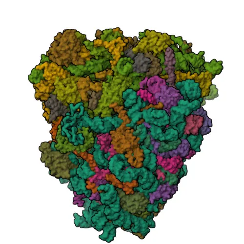
- RPL7A encodes the 60S ribosomal protein L7a, also designated as large ribosomal subunit protein eL8, and is known by aliases including PLA-X polypeptide and Surfeit locus protein 3.
- As a core component of the large ribosomal subunit, RPL7A is essential for ribosome biogenesis and the translation machinery, directly participating in cellular protein synthesis (PubMed:32669547, PubMed:23636399). Related references: PMID:32669547 PMID:23636399
- The ribosome, a large ribonucleoprotein complex, relies on subunits like RPL7A to decode mRNA into polypeptide chains, linking genotype to phenotype in all living cells (PubMed:32669547, PubMed:23636399). Related references: PMID:32669547 PMID:23636399
- RPL7A is localized predominantly in the cytoplasm, where it assembles into functional ribosomes, consistent with its role in translation.
- Post-translational modifications of RPL7A include acetylation, isopeptide bond formation, and ubiquitin conjugation, which may regulate ribosomal function or stability.
- RPL7A is associated with chromosomal rearrangements, such as those involving the surfeit locus, and is listed as a reference proteome component, underscoring its evolutionary conservation and clinical relevance.
Experimental Guidance and Technical Tips
- The immunogen encompasses the full-length human RPL7A protein (aa 1-266), suggesting the antibody may recognize both native and denatured forms in applications like WB and IF/ICC; consider performing a dilution series to determine the optimal titer for your specific assay.
- For Western blot, the predicted molecular weight is ~30 kDa; ensure proper gel percentage and transfer conditions for low molecular weight proteins, and validate loading controls such as GAPDH or β-actin.
- In immunofluorescence/ICC, characterize the subcellular localization (expected cytoplasmic) using appropriate fixation and permeabilization protocols; co-staining with organelle markers may help confirm specificity.
- Due to cross-reactivity with mouse and rat RPL7A, this antibody can be used for translational studies in common rodent models; consider verifying sequence homology if using other species.
- For ELISA, pair with a suitable secondary antibody and optimize coating antigen and detection conditions; recombinant RPL7A protein or cell lysates may serve as positive controls.
CamelBio: Your One-Stop Sourcing Bridge
CamelBio provides diagnostic manufacturers, labs, and research institutes with one-stop access to IVD raw materials, technical services, and consulting—covering every stage from concept to clinic. For ribosomal protein and translational research targets like RPL7A, we supply validated antibody pairs, optimized polyclonal antibodies, bulk ancillary reagents, and can assist with rare target sourcing. Contact us to streamline your next-generation diagnostic development pipeline.
REQUEST A QUOTE
Our professional team will reply to you within one business day. Please feel free to contact us!