Polyclonal Antibodies
Anti-RIPK1/RIP Rabbit Polyclonal Antibody for WB, ELISA - Q13546
Item Number : CM0025634
Price varies based on specs and customizations
- Application
- WB, ELISA
- Cross Reactivity
- Human, Mouse
- Protein Weight
- 75kDa

Shipping:
Contact us to get shipping details Enjoy On-time Dispatch Guarantee.
Why Choose Us
Easy ordering process, quality products, and dedicated support for your business success.
Core Product Specifications and Parameters
| Parameter | Value |
|---|---|
| Product Name | RIPK1/RIP Rabbit pAb |
| Remarks/Alias | RIP; RIP1; AIEFL; IMD57; RIP-1 |
| Species | Human |
| GeneID | 8737 |
| Immunogen | Recombinant fusion protein containing a sequence corresponding to amino acids 465-671 of human RIPK1/RIP (NP_003795.2) |
| Source | Rabbit |
| Category | Polyclonal Antibodies |
| Application | WB, ELISA |
| Cross Reactivity | Human, Mouse |
| SWISS | Q13546 |
| Protein Weight | 75kDa |
| Shipping | Ice bag |
Biological Background: RIPK1 Function and Localization
-
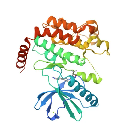
RIPK1 (Receptor-interacting serine/threonine-protein kinase 1), also known as RIP1, is a key regulator of TNF-mediated apoptosis, necroptosis, and inflammatory pathways. Related references: PMID:17703191, PMID:24144979, PMID:31827280
-
RIPK1 has a kinase-independent scaffold function in TNF-R1 signaling complex I, promoting cell survival by activating the canonical NF-kappa-B pathway; its kinase activity drives the assembly of death-inducing complexes IIa (apoptosis) and IIb (necroptosis). Related references: PMID:11101870, PMID:19524512, PMID:19524513
-
RIPK1 inhibits RIPK3-dependent necroptosis by facilitating FADD-mediated recruitment of CASP8, which cleaves RIPK1 and limits necroptosis. Related references: PMID:19524512, PMID:19524513, PMID:29440439
-
It promotes inflammatory responses by enhancing transcriptional production of pro-inflammatory cytokines such as interleukin-6 (IL6). Related references: PMID:31827280, PMID:31827281
-
RIPK1 phosphorylates RIPK3 (involved in reciprocal auto- and trans-phosphorylation) and DAB2IP at Ser-728, thereby activating the MAP3K5-JNK apoptotic cascade. Related references: PMID:19524513, PMID:15310755, PMID:17389591
-
Subcellular localization is mainly cytoplasmic and associated with the cell membrane, consistent with its role in receptor-proximal signaling events.
-
RIPK1 undergoes alternative splicing and is implicated in host-virus interactions, apoptosis, necrosis, and inflammatory responses, highlighting its diverse roles in cell fate decisions.
Experimental Guidance and Technical Tips
- The immunogen corresponds to amino acids 465–671 of human RIPK1, a region that is conserved between human and mouse, supporting the reported cross-reactivity.
- For Western blot (WB), consider using positive control lysates from human or mouse cell lines known to express RIPK1 (e.g., HeLa, Jurkat, or RAW 264.7). Expect a band near 75 kDa. Validate specificity in your sample system.
- For ELISA, both direct and sandwich formats can be explored; consider using recombinant RIPK1 protein or cell lysates for coating, and optimize detection conditions.
CamelBio: Your One-Stop Sourcing Bridge
CamelBio is your dedicated one-stop partner for IVD raw materials, offering a comprehensive portfolio including this high-quality rabbit polyclonal antibody against RIPK1. We support diagnostic manufacturers and researchers with reliable bulk sourcing of validated antibody pairs, optimized monoclonal/polyclonal antibodies, bulk ancillary reagents, and rare target raw materials across apoptosis, necroptosis, and inflammatory disease research. From concept to clinic, CamelBio streamlines your supply chain with expert technical support and consulting.
REQUEST A QUOTE
Our professional team will reply to you within one business day. Please feel free to contact us!