Monoclonal Antibodies
Anti-RIPK1/RIP Monoclonal Antibody for WB, IP, ELISA - Q13546
Item Number : CM0003896
Price varies based on specs and customizations
- Application
- WB, IP, ELISA
- Cross Reactivity
- Human
- Protein Weight
- 76kDa

Shipping:
Contact us to get shipping details Enjoy On-time Dispatch Guarantee.
Why Choose Us
Easy ordering process, quality products, and dedicated support for your business success.
Core Product Specifications and Parameters
| Parameter | Specification |
|---|---|
| Product Name | [KD Validated] RIPK1/RIP Rabbit mAb |
| Remarks/Alias | RIP; RIP1; AIEFL; IMD57; RIP-1; RIPK1/RIP |
| Species | Human |
| Gene ID (Human) | 8737 |
| Gene ID | 8737 |
| Immunogen | Recombinant protein |
| Source | Rabbit |
| Category | Monoclonal Antibodies |
| Application | WB, IP, ELISA |
| Cross Reactivity | Human |
| SWISS | Q13546 |
| Protein Weight | 76kDa |
| Shipping | Ice bag |
Biological Background: RIPK1 Function and Localization
- RIPK1, also known as RIP1 or cell death protein RIP, is a serine-threonine kinase that acts as a critical regulator of TNF-mediated apoptosis, necroptosis, and inflammatory signaling (PubMed:17703191, PubMed:24144979, PubMed:31827280). Related references: PMID:17703191, PMID:24144979, PMID:31827280
- It exhibits kinase activity-dependent functions in cell death and kinase-independent scaffold functions in cell survival, including activation of the canonical NF-kappa-B pathway (PubMed:11101870, PubMed:19524512, PubMed:19524513). Related references: PMID:11101870, PMID:19524512, PMID:19524513
- RIPK1 regulates assembly of two death-inducing complexes: complex IIa (RIPK1-FADD-CASP8) for apoptosis and complex IIb (RIPK1-RIPK3-MLKL) for necroptosis.
- It inhibits RIPK3-dependent necroptosis by recruiting CASP8 via FADD, which cleaves RIPK1, thus limiting TNF-induced necroptosis (PubMed:19524512, PubMed:19524513, PubMed:29440439). Related references: PMID:19524512, PMID:19524513, PMID:29440439
- RIPK1 is predominantly localized in the cytoplasm and at the cell membrane, consistent with its role in TNFR1 signal transduction.
- Key post-translational modifications include phosphorylation, ubiquitination (isopeptide bond), and proteolytic cleavage by CASP8, regulating its pro-death and pro-survival functions.
- It also promotes transcription of pro-inflammatory cytokines such as IL-6, linking cell death to immune responses (PubMed:31827280, PubMed:31827281). Related references: PMID:31827280, PMID:31827281
Experimental Guidance and Technical Tips
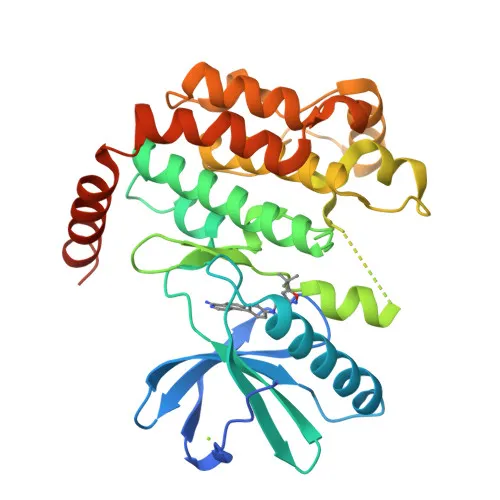
- The immunogen corresponds to amino acids 289-490 of human RIPK1, a region encompassing part of the kinase domain; this design may favor detection of both full-length and kinase-containing fragments.
- For Western blot, expect a band around 76 kDa. Consider including appropriate controls to account for cleavage products that can arise during apoptosis or necroptosis.
- In immunoprecipitation, validate antibody performance with known RIPK1 interactors such as TRADD or FADD under appropriate stimulation conditions (e.g., TNF treatment).
- ELISA experiments may be optimized for quantification of RIPK1 in cell lysates or tissue homogenates; cross-reactivity with other RIP family members should be assessed.
CamelBio: Your One-Stop Sourcing Bridge
CamelBio provides a streamlined sourcing channel for high-quality IVD raw materials, including validated antibody pairs, monoclonal antibodies, and bulk ancillary reagents for RIPK1-related research. We support diagnostic manufacturers in developing assays for cell death and inflammatory diseases, from early-stage R&D to clinical supply. With expertise in rare-target material sourcing, CamelBio helps you overcome procurement challenges and accelerate your IVD project timelines.
REQUEST A QUOTE
Our professional team will reply to you within one business day. Please feel free to contact us!