Polyclonal Antibodies
Anti-RIP3/RIPK3 Polyclonal Antibody for IHC-P and ELISA - Q9Y572
Item Number : CM0022111
Price varies based on specs and customizations
- Application
- IHC-P, ELISA
- Cross Reactivity
- Human
- Protein Weight
- 57kDa

Shipping:
Contact us to get shipping details Enjoy On-time Dispatch Guarantee.
Why Choose Us
Easy ordering process, quality products, and dedicated support for your business success.
Core Product Specifications and Parameters
| Parameter | Value |
|---|---|
| Product Name | RIP3 Rabbit pAb |
| Remarks/Alias | RIP3; RIPK3 |
| Species | Human |
| Gene ID | 11035 |
| Immunogen | A synthetic peptide corresponding to a sequence within amino acids 410-518 of human RIP3 (NP_006862.2). |
| Source | Rabbit |
| Category | Polyclonal Antibodies |
| Application | IHC-P, ELISA |
| Cross Reactivity | Human |
| SWISS | Q9Y572 |
| Protein Weight | 57kDa |
| Shipping | Ice bag |
Biological Background: RIP3 Function and Localization
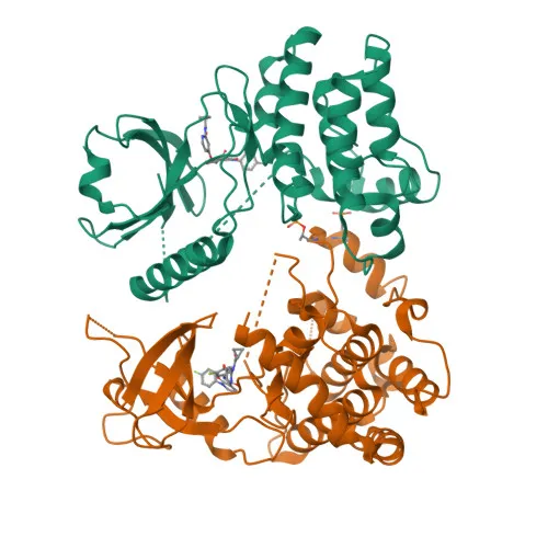
- RIP3 (Receptor-interacting serine/threonine-protein kinase 3), also known as RIPK3, is a serine/threonine kinase that activates both necroptosis and apoptosis, two parallel programmed cell death pathways. Related references: PMID:19524512, PMID:19524513, PMID:22265413
- Upon activation by ZBP1, RIPK3 forms a necrosis-inducing complex and phosphorylates MLKL, leading to plasma membrane permeabilization and necroptotic cell death. Related references: PMID:19524512, PMID:19524513, PMID:22265413
- RIPK3 also regulates extrinsic apoptosis through a RIPK1-, FADD-, and CASP8-dependent mechanism that is independent of MLKL and its own kinase activity.
- The protein localizes predominantly to the cytosol but can also be found in the nucleus, where it mediates nuclear necroptosis in response to viral infections such as orthomyxoviruses.
- RIPK3 is highly expressed in the pancreas and detected at lower levels in heart, placenta, lung, and kidney; its expression is significantly increased in colon and lung cancers.
- In certain cell types, RIPK3 restricts viral replication through death-independent pathways, for example by upregulating ACOD1/IRG1 and itaconate synthesis upon Zika virus infection.
- RIPK3 binds to and enhances the activity of metabolic enzymes GLUL, GLUD1, and PYGL, potentially stimulating the tricarboxylic acid cycle, oxidative phosphorylation, and ROS production. Related references: PMID:19498109
- The kinase undergoes phosphorylation, ubiquitination, and isopeptide bond formation, and is subject to alternative splicing, contributing to functional diversity.
Experimental Guidance and Technical Tips
- This polyclonal antibody is raised against a synthetic peptide spanning amino acids 410-518 (C-terminal region) of human RIP3. It is expected to recognize full-length RIP3 and may also detect splice variants containing this epitope.
- For IHC-P, consider optimizing antigen retrieval conditions (e.g., heat-mediated citrate or EDTA buffers) and validate staining specificity using appropriate tissue controls (positive and negative) and, if possible, pre-absorption with the immunogen peptide.
- In ELISA, ensure the coating antigen corresponds to the immunogen region or use recombinant full-length protein. Check for batch-to-batch consistency by titrating the antibody against a known standard.
- As a polyclonal reagent, lot-to-lot variability may occur; include internal referencing when comparing results across experiments.
CamelBio: Your One-Stop Sourcing Bridge
CamelBio provides a comprehensive portfolio of IVD raw materials, including validated antibody pairs and optimized polyclonal antibodies like this anti-RIP3 product, to accelerate necroptosis and cell death research. Our platform streamlines procurement from discovery to clinical application, supplying manufacturers and laboratories with reliable reagents and technical guidance. Whether you need bulk ancillary reagents or assistance sourcing rare-target antibodies, CamelBio is your single partner for seamless IVD development.
REQUEST A QUOTE
Our professional team will reply to you within one business day. Please feel free to contact us!