Polyclonal Antibodies
Anti-RIP3 Rabbit Polyclonal Antibody for IHC-P, IF/ICC, ELISA - Q9QZL0
Item Number : CM0020566
Price varies based on specs and customizations
- Application
- IHC-P, IF/ICC, ELISA
- Cross Reactivity
- Human, Mouse, Rat
- Protein Weight
- 53kDa

Shipping:
Contact us to get shipping details Enjoy On-time Dispatch Guarantee.
Why Choose Us
Easy ordering process, quality products, and dedicated support for your business success.
Core Product Specifications and Parameters
| Parameter | Value |
|---|---|
| Product Name | RIP3 Rabbit pAb |
| Remarks/Alias | Rip3; 2610528K09Rik; RIP3 |
| Species | Mouse |
| GeneID (Human) | 11035 |
| GeneID | 56532 |
| Immunogen | Synthetic peptide corresponding to a sequence within amino acids 320-420 of mouse RIP3 (NP_064339.2) |
| Source | Rabbit |
| Category | Polyclonal Antibodies |
| Application | IHC-P, IF/ICC, ELISA |
| Cross Reactivity | Human, Mouse, Rat |
| SWISS | Q9QZL0 |
| Protein Weight | 53kDa |
| Shipping | Ice bag |
Biological Background: RIP3 Function and Localization
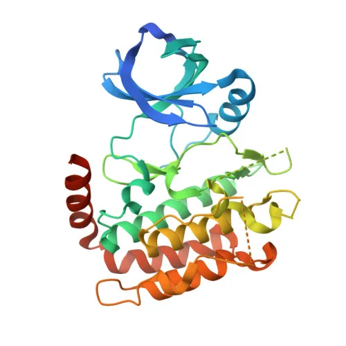
- RIP3 (Ripk3), also known as RIP-like protein kinase 3 or receptor-interacting protein 3, is a serine/threonine kinase that activates both necroptosis and apoptosis (PubMed:27321907, PubMed:27746097, PubMed:27917412). Related references: PMID:27321907, PMID:27746097, PMID:27917412
- Necroptosis is triggered by RIPK3 following activation by ZBP1 (PubMed:19590578, PubMed:22423968, PubMed:24012422). Related references: PMID:19590578, PMID:22423968, PMID:24012422
- Activated RIPK3 forms a necrosis-inducing complex and phosphorylates MLKL, leading to plasma membrane damage and calcium influx (PubMed:24813849, PubMed:24813850, PubMed:27321907). Related references: PMID:24813849, PMID:24813850, PMID:27321907
- In the nucleus, RIPK3 can mediate necroptosis in response to orthomyxovirus infection by phosphorylating MLKL, causing nuclear envelope disruption (PubMed:32200799, PubMed:32296175). Related references: PMID:32200799, PMID:32296175
- RIPK3 also regulates apoptosis in a RIPK1-, FADD-, and CASP8-dependent manner, independent of MLKL and its kinase activity (PubMed:27321907). Related references: PMID:27321907
- Apart from cell death, RIPK3 restricts viral replication via a death-independent pathway by upregulating ACOD1/IRG1 and itaconate production in neurons (PubMed:30635240). Related references: PMID:30635240
- RIPK3 binds to and enhances activity of metabolic enzymes GLUL, GLUD1, and PYGL, potentially stimulating ROS production (By similarity).
- Subcellularly, RIPK3 localizes to the cytoplasm, cytosol, and nucleus.
- Tissue expression includes embryo and adult spleen, liver, testis, heart, brain, and lung.
Experimental Guidance and Technical Tips
- The immunogen is a synthetic peptide corresponding to amino acids 320-420 of mouse RIP3; the antibody may recognize the C-terminal region. For IHC-P, consider using tissue sections known to express RIP3 (e.g., spleen, liver) and validate with appropriate positive and negative controls.
- For IF/ICC, fixation and permeabilization conditions should be optimized to preserve epitope accessibility; test a range of conditions to determine optimal staining.
- In ELISA, this polyclonal antibody can be used as a capture or detection reagent; titration and pairing with a matched secondary antibody are recommended.
- As a polyclonal antibody, lot-to-lot consistency should be verified, and each new batch validated in the intended application.
CamelBio: Your One-Stop Sourcing Bridge
CamelBio offers a comprehensive procurement solution for IVD raw materials, serving diagnostic developers and research labs from concept to clinic. This anti-RIP3 polyclonal antibody supports necroptosis, apoptosis, and cell death research, and we can further supply validated antibody pairs, optimized monoclonal antibodies, bulk ancillary reagents, and rare target sourcing to accelerate your assay development.
Product Datasheet
Anti-RIP3 Rabbit Polyclonal Antibody for IHC-P, IF/ICC, ELISA - Q9QZL0
REQUEST A QUOTE
Our professional team will reply to you within one business day. Please feel free to contact us!