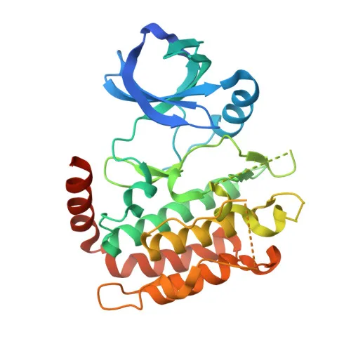
Monoclonal Antibodies
Anti-RIP3 Rabbit Monoclonal Antibody for WB, IF/ICC, ELISA - Q9QZL0
Item Number : CM0005582
Price varies based on specs and customizations
- Application
- WB, IF/ICC, ELISA
- Cross Reactivity
- Mouse
- Protein Weight
- 63kDa

Shipping:
Contact us to get shipping details Enjoy On-time Dispatch Guarantee.
Why Choose Us
Easy ordering process, quality products, and dedicated support for your business success.
Core Product Specifications and Parameters
| Parameter | Value |
|---|---|
| Product Name | RIP3 Rabbit mAb |
| Remarks/Alias | Rip3; 2610528K09Rik |
| Species | Mouse |
| GeneID (Human) | 11035 |
| GeneID | 56532 |
| Immunogen | Synthetic peptide|A synthetic peptide corresponding to a sequence within amino acids 320-420 of mouse RIP3 (NP_064339.2). |
| Source | Rabbit |
| Category | Monoclonal Antibodies |
| Application | WB, IF/ICC, ELISA |
| Cross Reactivity | Mouse |
| SWISS | Q9QZL0 |
| Protein Weight | 63kDa |
| Shipping | Ice bag |
Biological Background: RIP3/RIPK3 Function and Localization
- Receptor-interacting serine/threonine-protein kinase 3 (RIPK3, also known as RIP3) is a key mediator of necroptosis and apoptosis, two parallel programmed cell death pathways (PubMed:27321907, PubMed:27746097, PubMed:27917412). Related references: PMID:27321907, PMID:27746097, PMID:27917412
- Necroptosis is triggered by activation of RIPK3 by ZBP1, leading to formation of a necrosis-inducing complex and phosphorylation of MLKL, which translocates to the plasma membrane to cause membrane damage and calcium influx (PubMed:19590578, PubMed:22423968, PubMed:24012422, PubMed:24813849, PubMed:24813850). Related references: PMID:19590578, PMID:22423968, PMID:24012422, PMID:24813849, PMID:24813850
- In the nucleus, upon orthomyxovirus infection, ZBP1 senses Z-RNA and activates RIPK3 to phosphorylate MLKL, disrupting the nuclear envelope and leaking DNA into the cytosol (PubMed:32200799, PubMed:32296175). Related references: PMID:32200799, PMID:32296175
- RIPK3 can also restrict viral replication through cell death-independent mechanisms, such as upregulating ACOD1/IRG1 and itaconate production to inhibit succinate dehydrogenase and suppress viral genome replication (PubMed:30635240). Related references: PMID:30635240
- RIPK3 is predominantly cytosolic but can also localize to the nucleus, and is expressed in embryo and adult tissues including spleen, liver, testis, heart, brain, and lung.
- The protein is regulated by phosphorylation, methylation, and ubiquitin-like conjugation, and it can also influence cellular metabolism by interacting with metabolic enzymes like GLUL, GLUD1, and PYGL to stimulate the TCA cycle and oxidative phosphorylation, potentially increasing ROS production.
Experimental Guidance and Technical Tips
- The immunogen corresponds to amino acids 320–420 within the kinase domain of mouse RIP3, suggesting the antibody may recognize epitopes in this functional region.
- For western blot, RIP3 has an observed molecular weight of ~63 kDa; consider loading appropriate positive controls such as mouse spleen or liver lysates.
- For immunofluorescence/immunocytochemistry, RIP3 localizes to the cytoplasm and nucleus; include appropriate fixation and permeabilization steps.
- In ELISA, the antibody can be used for detection of recombinant or endogenous RIP3; optimization of coating and detection conditions is recommended.
- Cross-reactivity has been validated in mouse; cross-reactivity in other species is not guaranteed and should be experimentally verified.
CamelBio: Your One-Stop Sourcing Bridge
CamelBio provides diagnostic manufacturers and research laboratories with a comprehensive portfolio of IVD raw materials, including validated antibodies for necroptosis and cell death research. With expertise in monoclonal antibody development and bulk reagent supply, we support your projects from concept to clinic. Contact us for customized solutions, including antibody pairs and rare-target reagent sourcing.
REQUEST A QUOTE
Our professional team will reply to you within one business day. Please feel free to contact us!