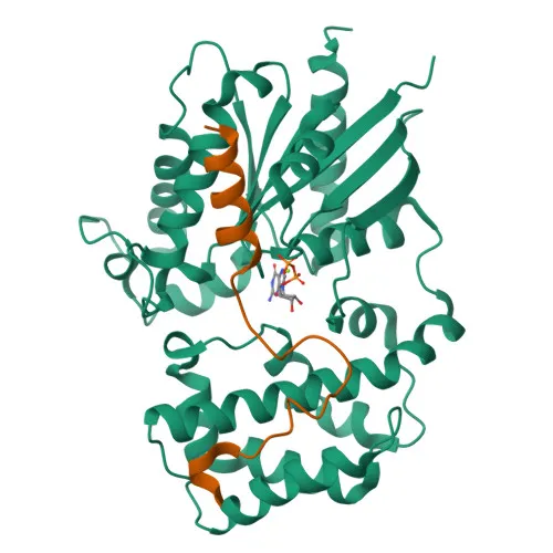
Polyclonal Antibodies
Anti-RGS14 Polyclonal Antibody for WB, IHC-P, ELISA - O43566
Item Number : CM0030142
Price varies based on specs and customizations
- Application
- WB, IHC-P, ELISA
- Cross Reactivity
- Human, Mouse, Rat
- Protein Weight
- 61kDa

Shipping:
Contact us to get shipping details Enjoy On-time Dispatch Guarantee.
Why Choose Us
Easy ordering process, quality products, and dedicated support for your business success.
Core Product Specifications and Parameters
| Parameter | Value |
|---|---|
| Product Name | RGS14 Rabbit pAb |
| Remarks/Alias | RGS14 |
| Species | Human |
| GeneID (Human) | 10636 |
| GeneID | 10636 |
| Immunogen | Recombinant fusion protein containing a sequence corresponding to amino acids 230-510 of human RGS14 (NP_006471.2) |
| Source | Rabbit |
| Category | Polyclonal Antibodies |
| Application | WB, IHC-P, ELISA |
| Cross Reactivity | Human, Mouse, Rat |
| SWISS | O43566 |
| Protein Weight | 61kDa |
| Shipping | Ice bag |
Biological Background: RGS14 Function and Localization
- Regulator of G protein signaling 14 (RGS14) is a multifunctional protein that modulates G protein-coupled receptor (GPCR) signaling by acting as a GTPase-activating protein (GAP) and a GDP-dissociation inhibitor (GDI) for specific G alpha subunits.
- It inhibits signal transduction by increasing GTPase activity of G(i) alpha subunits GNAI0, GNAI2, and GNAI3, and has GDI activity on GNAI1 and GNAI3, but not on GNAI2 or G(o)-alpha.
- RGS14 integrates G protein and Ras/Raf MAP kinase signaling pathways; it inhibits PDGF-stimulated ERK1/ERK2 phosphorylation in a HRAS-dependent manner that is reversed by GNAI1.
- It plays a role in cell division by positively modulating microtubule polymerization and spindle organization through a G(i)-alpha-dependent mechanism.
- The protein is required for nerve growth factor (NGF)-mediated neurite outgrowth and is involved in visual memory processing capacity and hippocampal-based learning and memory.
- Subcellular localization is diverse: found in the nucleus (including PML bodies), cytoplasm, cell membrane, centrosome, spindle, spindle pole, dendrites, dendritic spines, and postsynaptic density.
- The gene undergoes alternative splicing, and the protein is classified under keywords including phosphoprotein, signal transduction inhibitor, synapse, and cell cycle.
Experimental Guidance and Technical Tips
- The immunogen was a recombinant protein covering amino acids 230-510 of human RGS14; this region may be conserved across species, supporting the observed cross-reactivity with mouse and rat.
- For western blotting (WB), the predicted molecular weight is approximately 61 kDa; consider running appropriate positive controls (e.g., human, mouse, or rat cell lysates) and optimizing the primary antibody dilution.
- In immunohistochemistry (IHC-P), use standard antigen retrieval methods and optimize the detection protocol; note the wide tissue distribution of RGS14, including brain, where it localizes to dendritic spines.
- For ELISA, the antibody may be suitable for capture or detection; validate against the immunogen or full-length protein in your assay system.
- Always include negative controls (e.g., secondary antibody alone) and, if possible, knockdown or knockout lysates to confirm specificity.
CamelBio: Your One-Stop Sourcing Bridge
CamelBio serves as a comprehensive sourcing partner for IVD raw materials, technical services, and consulting—guiding projects from concept to clinic. For research into GPCR signaling, cell division, and neurological memory studies, we offer validated antibody pairs, optimized monoclonal and polyclonal antibodies, bulk ancillary reagents, and dedicated rare-target raw-material sourcing to support your diagnostic development and preclinical programs.
REQUEST A QUOTE
Our professional team will reply to you within one business day. Please feel free to contact us!