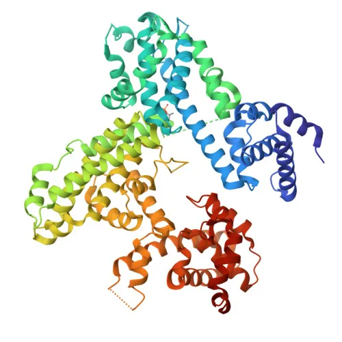
Monoclonal Antibodies
Anti-RB Rabbit mAb for WB, IP, IHC-P, ELISA - P06400
Item Number : CM0008503
Price varies based on specs and customizations
- Application
- WB, IP, IHC-P, ELISA
- Cross Reactivity
- Human
- Protein Weight
- 106kDa

Shipping:
Contact us to get shipping details Enjoy On-time Dispatch Guarantee.
Why Choose Us
Easy ordering process, quality products, and dedicated support for your business success.
Core Product Specifications and Parameters
| Parameter | Value |
|---|---|
| Product Name | RB Rabbit mAb |
| Remarks/Alias | RB; pRb; OSRC; pp110; p105-Rb; PPP1R130; p110-RB1 |
| Species | Human |
| Gene ID (Human) | 5925 |
| Gene ID | 5925 |
| Immunogen | Recombinant protein (Recombinant fusion protein containing a sequence corresponding to amino acids 701-928 of human RB - NP_000312.2) |
| Source | Rabbit |
| Category | Monoclonal Antibodies |
| Application | WB, IP, IHC-P, ELISA |
| Cross Reactivity | Human |
| SWISS | P06400 |
| Protein Weight | 106kDa |
| Shipping | Ice bag |
Biological Background: RB Function and Localization
- Retinoblastoma-associated protein (RB1) is a tumor suppressor encoded by the RB1 gene, also known as pRb, pp110, p105-Rb, or p110-RB1. The canonical isoform has a molecular weight of approximately 106 kDa.
- RB1 is a key regulator of the G1/S cell cycle transition: hypophosphorylated RB1 binds and represses E2F transcription factors, blocking expression of genes required for S-phase entry. Cyclin/CDK-dependent phosphorylation releases E2F, triggering cell cycle progression. RB1 also promotes the G0–G1 transition through CDK3/cyclin-C-mediated phosphorylation. Related references: PMID:10499802 PMID:15084261
- RB1 maintains overall chromatin structure and contributes to heterochromatin formation by recruiting histone methyltransferases (SUV39H1, KMT5B, KMT5C) and controlling H4 Lys-20 trimethylation.
- Subcellular localization is predominantly nuclear; however, RB1 can also shuttle to the cytoplasm, where it interacts with various factors during specific cell cycle phases or apoptotic signaling.
- During viral infection, oncoproteins from DNA tumor viruses (e.g., SV40 large T antigen, HPV E7, adenovirus E1A) bind RB1 and disrupt the RB1-E2F repressor complex, leading to uncontrolled cell proliferation.
- RB1 is expressed in the retina and in foreskin keratinocytes, consistent with its tissue-specific tumor suppressive roles. Related references: PMID:20940255
- The protein is subject to extensive post-translational modifications, including phosphorylation, acetylation, and methylation, which finely tune its activity and interactions.
Experimental Guidance and Technical Tips
- The immunogen was designed against the C-terminal region (amino acids 701–928) of human RB1; this region covers part of the pocket domain and the C-terminal domain, which are critical for protein interactions.
- For Western blot (WB), a band of approximately 106 kDa is expected; consider including phosphatase inhibitors in lysis buffers to preserve the endogenous phosphorylation state.
- The antibody has been tested in IP, IHC-P, and ELISA; each application may require distinct sample preparation and validation in the relevant sample system.
- Since cross-reactivity is reported only for human, verify reactivity in other species if multi-species experiments are planned.
CamelBio: Your One-Stop Sourcing Bridge
CamelBio supplies this RB1 monoclonal antibody as part of a comprehensive IVD raw-material portfolio for oncology and cell cycle research. Researchers and diagnostic manufacturers can access validated antibody pairs, optimized recombinant proteins, bulk ancillary reagents, and rare-target sourcing—all conveniently consolidated. From concept to clinic, CamelBio bridges the gap to help you accelerate assay development.
REQUEST A QUOTE
Our professional team will reply to you within one business day. Please feel free to contact us!