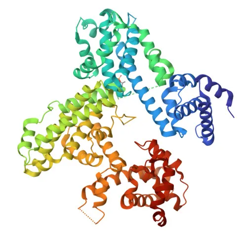
Polyclonal Antibodies
Anti-RB Polyclonal Antibody for WB, IF/ICC, ELISA - P06400
Item Number : CM0021984
Price varies based on specs and customizations
- Application
- WB, IF/ICC, ELISA
- Cross Reactivity
- Human
- Protein Weight
- 106 kDa

Shipping:
Contact us to get shipping details Enjoy On-time Dispatch Guarantee.
Why Choose Us
Easy ordering process, quality products, and dedicated support for your business success.
Core Product Specifications and Parameters
| Parameter | Specification |
|---|---|
| Product Name | RB Rabbit pAb |
| Remarks/Alias | RB; pRb; OSRC; pp110; p105-Rb; PPP1R130; p110-RB1 |
| Species | Human |
| Gene ID (Human) | 5925 |
| Immunogen | Recombinant fusion protein containing a sequence corresponding to amino acids 701-928 of human RB (NP_000312.2) |
| Source | Rabbit |
| Category | Polyclonal Antibodies |
| Application | WB, IF/ICC, ELISA |
| Cross Reactivity | Human |
| SWISS | P06400 |
| Protein Weight | 106 kDa |
| Shipping | Ice bag |
Biological Background: RB Function and Localization
-
RB (retinoblastoma-associated protein) is a tumor suppressor and key regulator of the G1/S cell cycle transition. The hypophosphorylated form binds E2F transcription factors, repressing transcription of genes required for S-phase entry. Cyclin/CDK-dependent phosphorylation releases E2Fs, triggering cell cycle progression (PubMed:10499802). RB also promotes the G0-G1 transition via phosphorylation by CDK3/cyclin-C (PubMed:15084261).
Related references: PMID:10499802 PMID:15084261
-
Directly involved in heterochromatin formation by stabilizing histone methylation and recruiting histone methyltransferases SUV39H1, KMT5B and KMT5C. Controls histone H4 lysine-20 trimethylation, leading to epigenetic transcriptional repression.
-
Mediates transcriptional repression by SMARCA4/BRG1 via recruitment of a histone deacetylase (HDAC) complex to the c-FOS promoter. In resting neurons, a phospho-RB1-HDAC1 repressor complex inhibits c-FOS transcription; calcium influx dephosphorylates RB1 via calcineurin, releasing the repression.
-
Subcellular localization includes the nucleus and cytoplasm.
-
Expressed in the retina and foreskin keratinocytes (at protein level) (PubMed:20940255).
Related references: PMID:20940255
-
Subject to key post‑translational modifications: acetylation, methylation, and extensive phosphorylation that regulate its activity and interactions.
-
During viral infection, SV40 large T antigen, HPV E7 protein, or adenovirus E1A protein bind RB1, disrupting the RB1‑E2F1 complex and abrogating its tumor suppressor function.
Experimental Guidance and Technical Tips
- The immunogen corresponds to the C‑terminal region (aa 701‑928), which is well‑conserved and likely to generate reactivity against full‑length RB (~106 kDa).
- For Western blot, use a mid‑range protein ladder to resolve the 106 kDa band clearly. Nuclear extracts are recommended for optimal detection of endogenous RB. Validate the antibody in relevant human cell lines (e.g., those with known RB status) and consider a blocking peptide control if background is observed.
- In IF/ICC, optimize fixation and permeabilization conditions; methanol fixation may better preserve nuclear RB epitopes. For ELISA, the recombinant immunogen can serve as a positive control.
- As a polyclonal antibody raised in rabbit, lot‑to‑lot consistency may vary. Include appropriate controls and validate each new lot.
CamelBio: Your One‑Stop Sourcing Bridge
CamelBio provides diagnostic manufacturers, research laboratories, and institutes with a comprehensive portfolio of IVD‑grade antibodies and raw materials. Our RB polyclonal antibody supports fundamental research in cell cycle control, tumor biology, and host‑virus interactions. Beyond this product, we offer validated antibody pairs, optimized monoclonal and polyclonal antibodies, bulk ancillary reagents, and assistance in sourcing rare‑target raw materials to accelerate your assay development from concept to clinic.
REQUEST A QUOTE
Our professional team will reply to you within one business day. Please feel free to contact us!